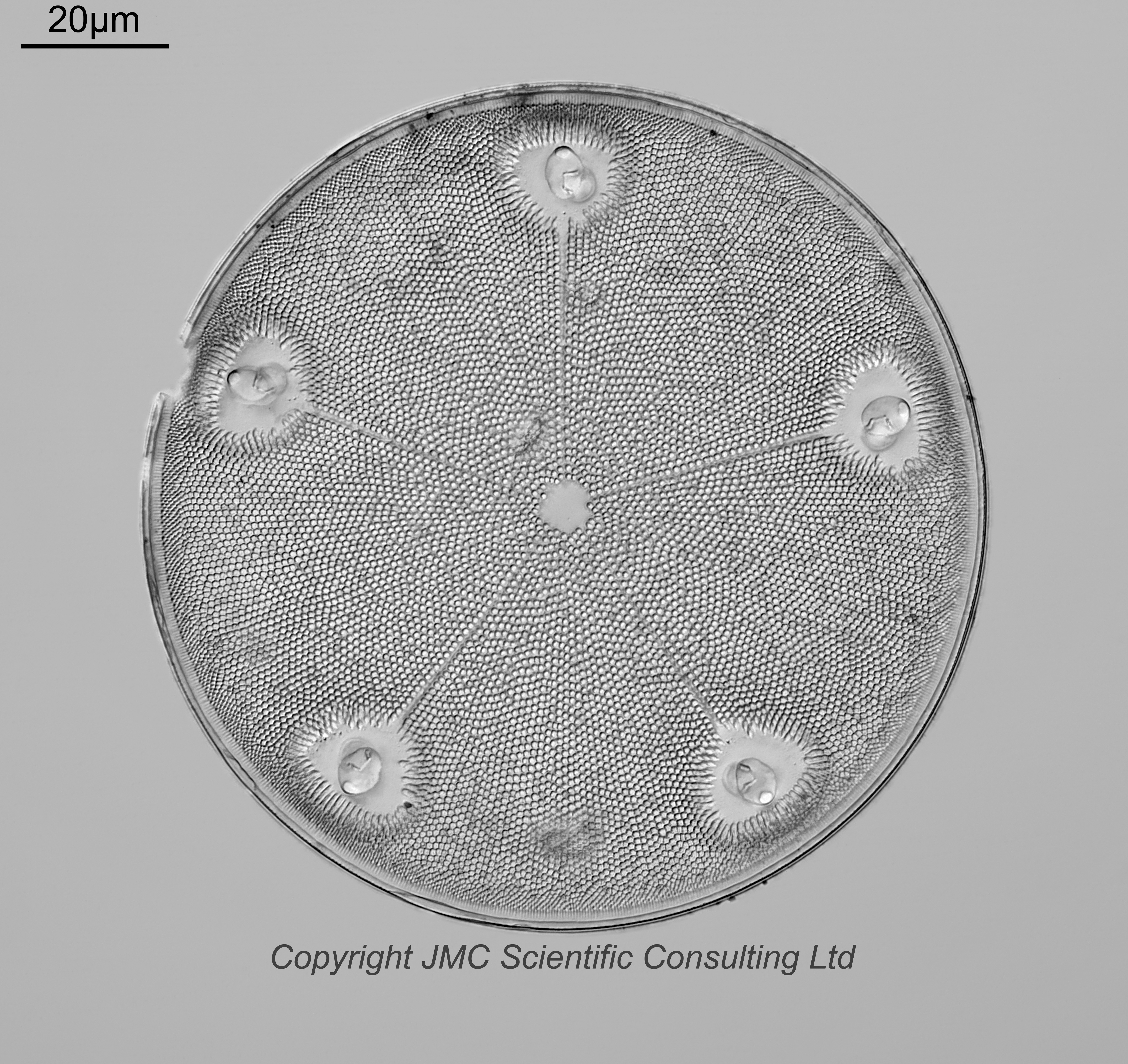
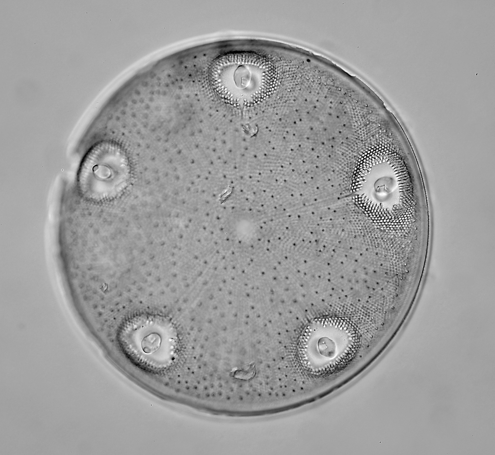
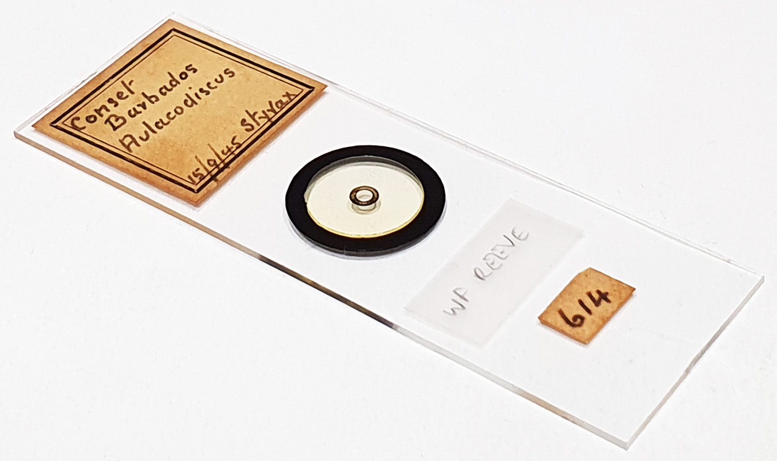
Aulacodiscus sp. (perhaps A. barbadensis ?) from Conset, Barbados. Single example on the slide. Dated 15/9/1945 and mounted in Styrax. No makers name but based on style and handwriting I think this is by WF Reeve. Olympus BHB microscope using 450nm LED light. 63x Leitz Pl Apo 1.4 objective, oil immersion. Olympus Aplanat Achromat condenser, oil immersion, oblique lighting. 2.5x Nikon CF PL photoeyepiece. Monochrome converted Nikon d850 camera. 39 images stacked in Zerene.
Ok, the name. In Oamaru there is a 4 process diatom which looks quite similar to this in terms of the shape of the processes and the hyaline region around them. This is called Aulacodiscus barbadensis. Based on the name (barabensis) I would certainly not be surprised to see this in Barbados. It is described in: Pritchard, A. (1861). A history of infusoria, including the Desmidiaceae and Diatomaceae, British and foreign. Fourth edition enlarged and revised by J. T. Arlidge, M.B., B.A. Lond.; W. Archer, Esq.; J. Ralfs, M.R.C.S.L.; W. C. Williamson, Esq., F.R.S., and the author. pp. [1]-xii, [1]-968, 40 pls [I-XV]. Page 939. London: Whittaker and Co.. The one described here has 3 processes, however no image is given. We have a 4 process one with an image (from Oamaru), and a description of a 3 process one from Barbados. I think this one is a 5 process example of A. barbadensis (a variant perhaps or just 5 processes instead of 3). What’s even more intriguing, on a 123 form slide alsofrom Conset, mounted by Barber (see here) there is something which looks pretty much identical to this one. At the time I just left it as Aulacodiscus sp. but now I am pretty confident that it is a 5 process A. barbadensis. On that one there are very visible dark dots across the face of the diatom. They are less visible here in the final stack, however they do show up in the individual frames. I suspect the strongly defined polygonal shapes on the surface of the diatom overrode these dark dots in the stacking. Also, this one is viewed from the top, while the other one is viewed from below. As those dark dots are on the top surface of the diatom, this means that with the other one they become visible ‘after’ the polygons during the stacking process. This would also account for them being more visible in that other one. One final observation, the diatom looks quite flat at first glance, but there is actually a slight circular depression between each of the processes. These depressions are only a few microns deep but showed up while I was focusing and capturing the individual images.