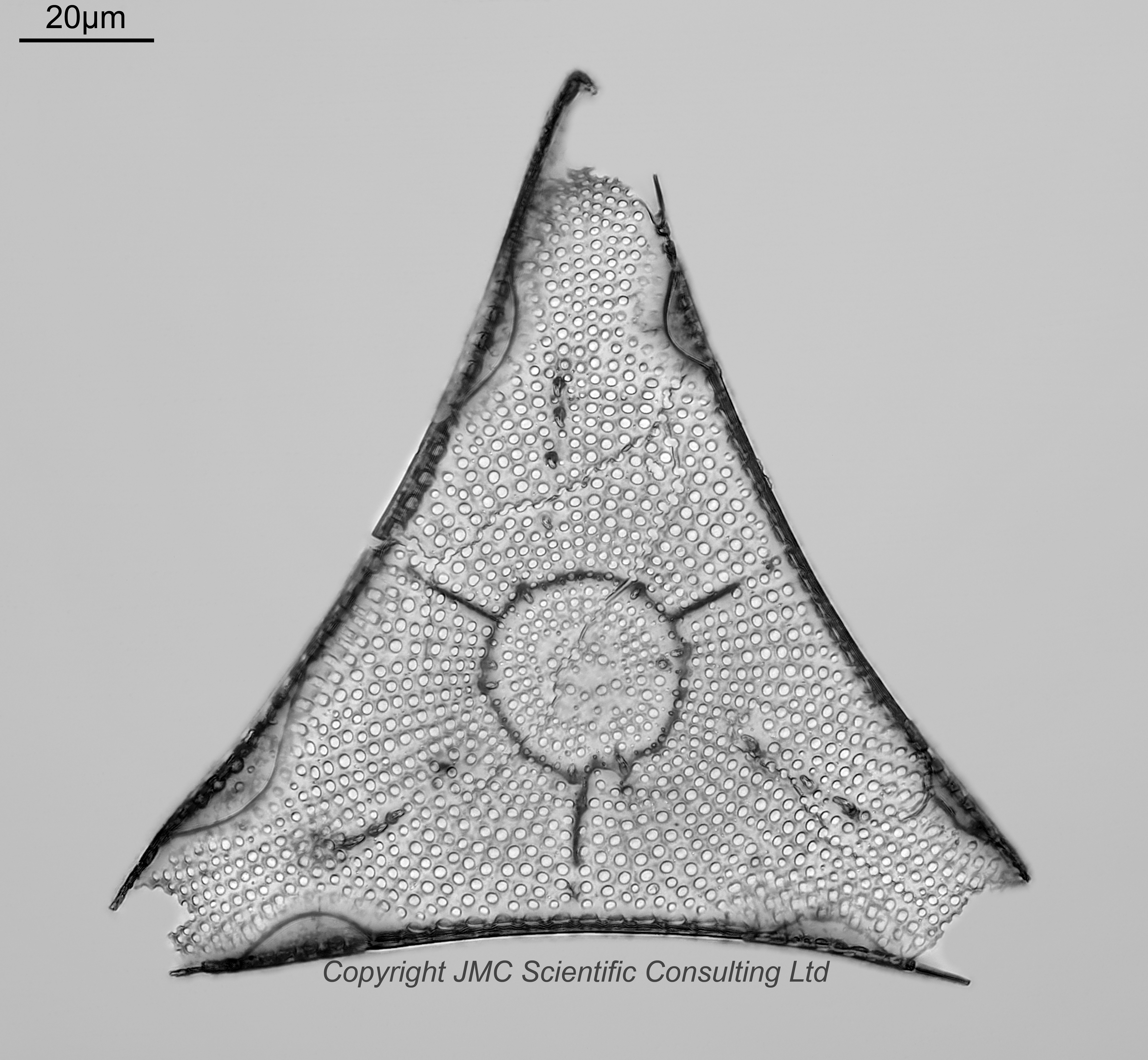
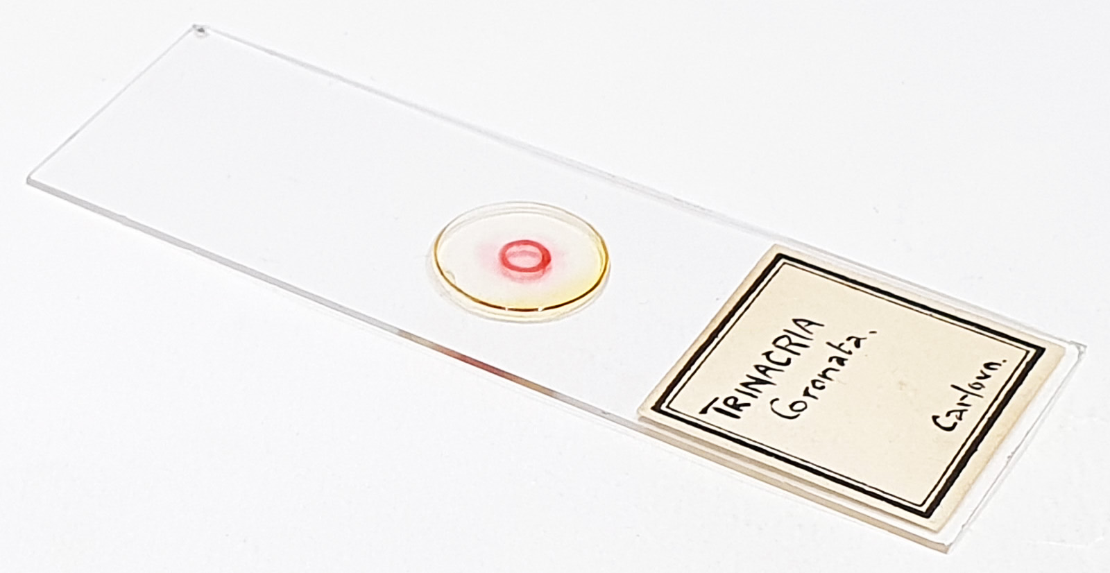
Trinacria coronata from Carlova. Incomplete, and rim facing the coverslip (underside view). Single example on the slide. No makers name. Olympus BHB microscope using 450nm LED light. 63x Leitz Pl Apo 1.4 objective, oil immersion. Olympus Aplanat Achromat condenser, oil immersion, oblique lighting. 2.5x Nikon CF PL photoeyepiece. Monochrome converted Nikon d850 camera. 61 images stacked in Zerene (Pmax). Some adhesive to the coverslip visible towards the apices. I wonder what makes the dark colour circle and linear features that are so apparent on examples of this species?
I recently acquired about 60 slides by this maker, so if you want to see others by them, search for AAAAA in the Search option at the top of the page, and I’ll include this in each of the pages for them.