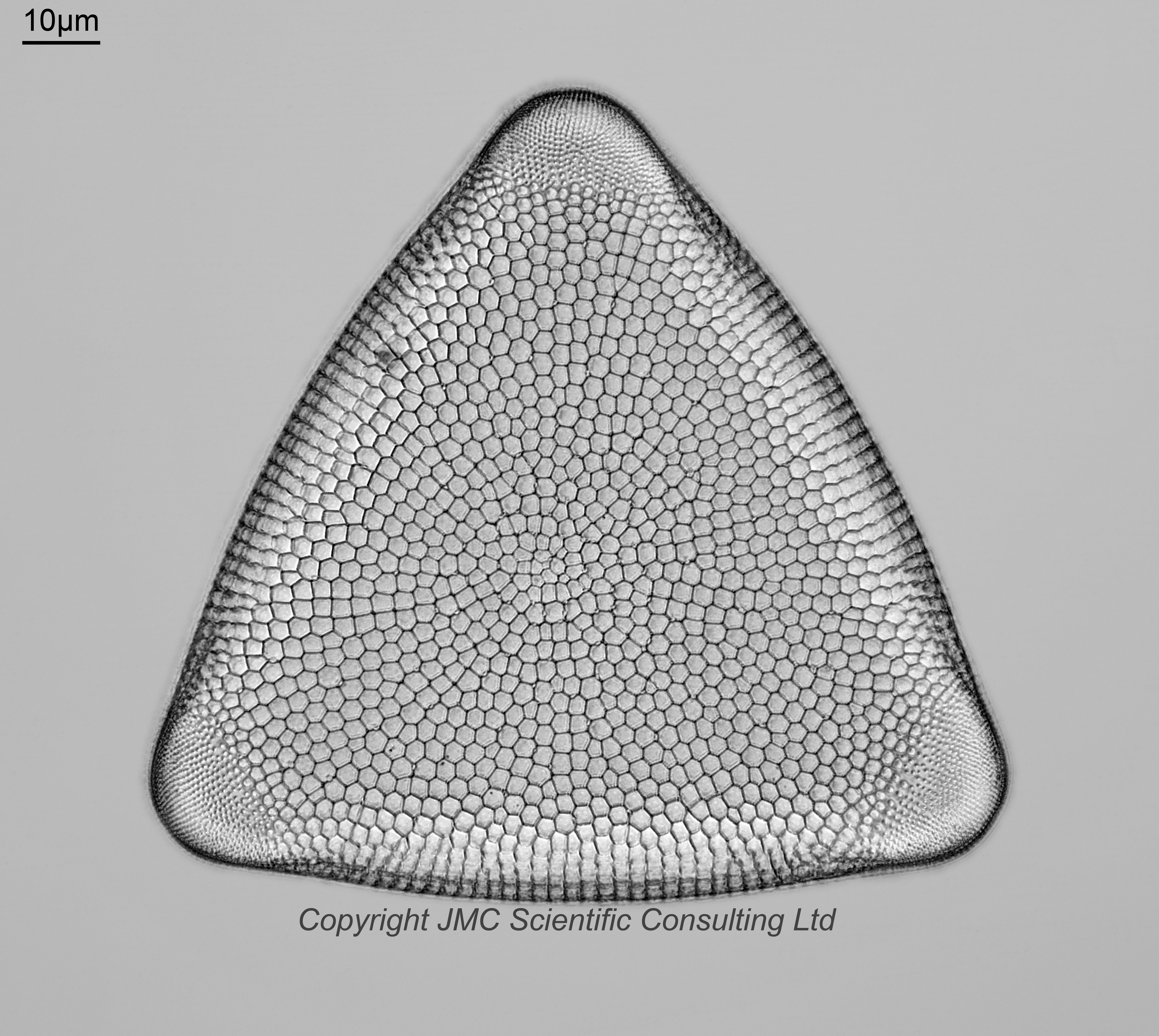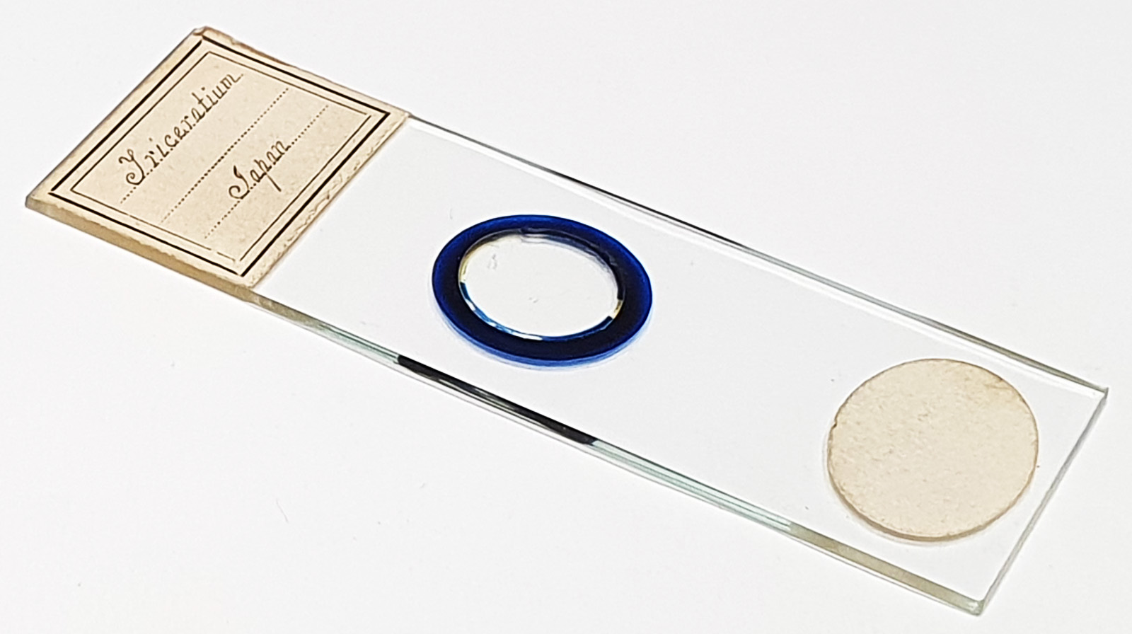


Triceratium sp. from Japan. Single example on the slide. No makers name, but the handwriting would have this as being prepared by Frederick Marshall. Olympus BHB microscope using 450nm LED light. 63x Leitz Pl Apo 1.4 objective, oil immersion. Olympus Aplanat Achromat condenser, oil immersion, slightly oblique lighting. 2.5x Nikon CF PL photoeyepiece. Monochrome converted Nikon d850 camera. 40 images stacked in Zerene (Pmax).
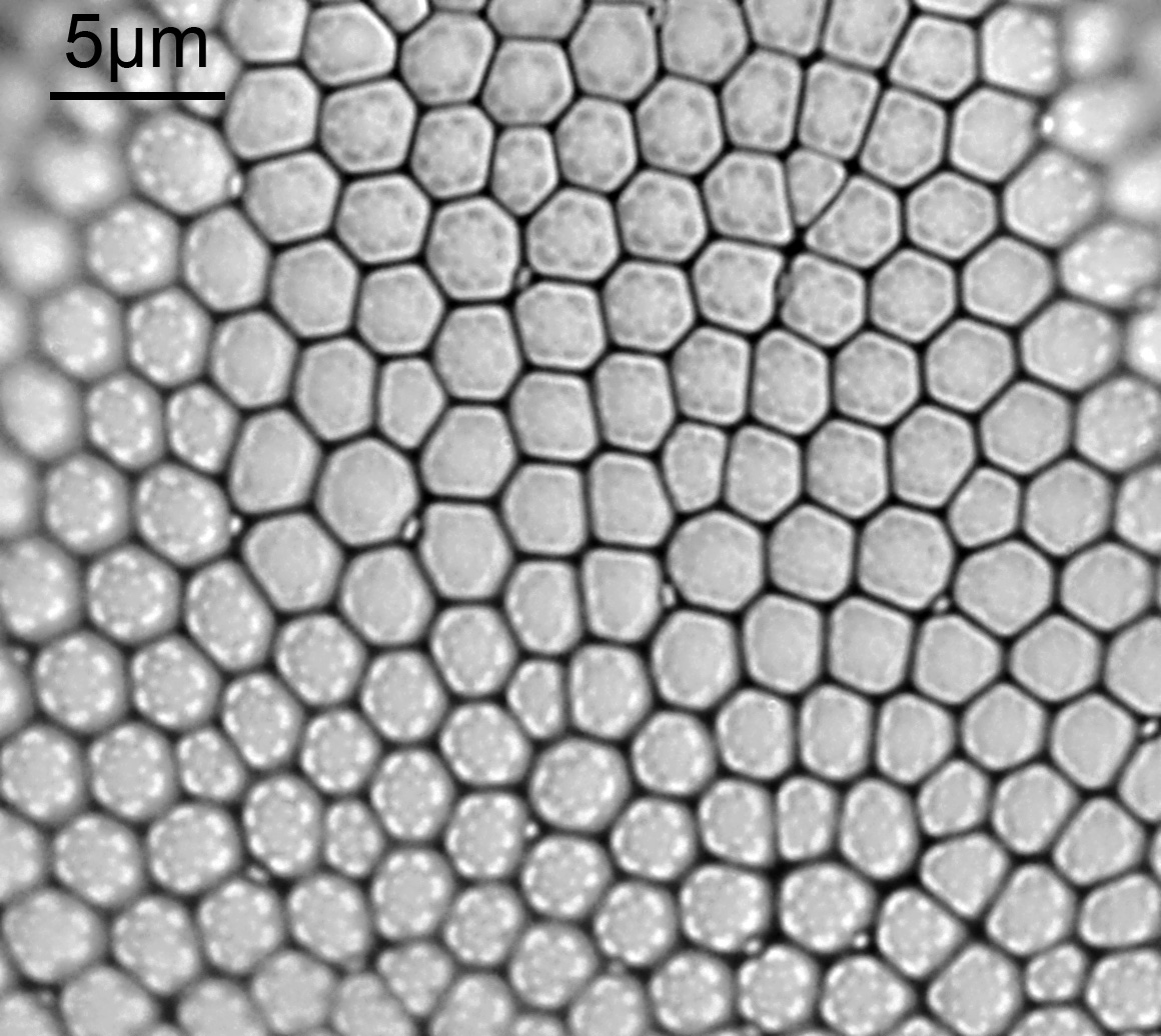
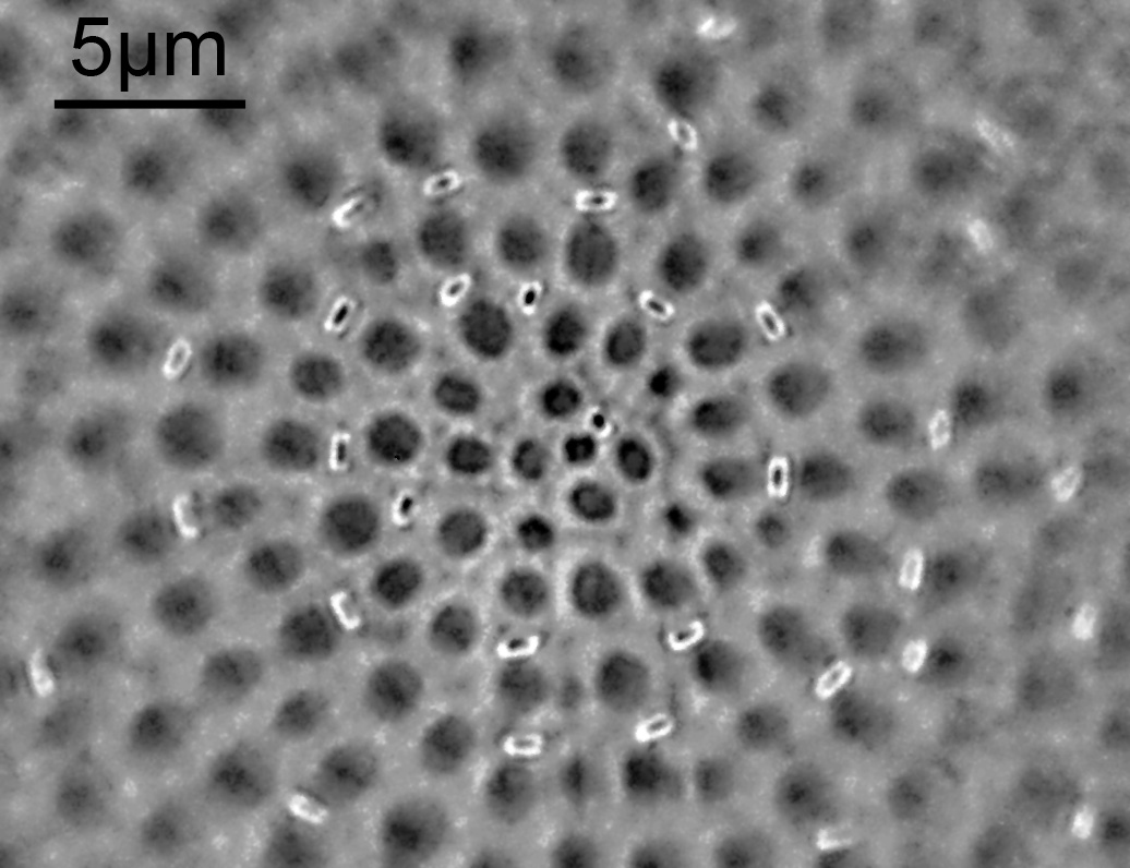
The original frames for the stack were relatively low contrast, even with the oblique lighting, and some of the structures present at different depths on the diatom were lost as a result of the stacking process. This can be seen when looking at crops from two of the original frames, which show different features. The images are not co-located, and have had their contrast boosted to show the features. In the second image I am presuming the bright features are the tubes of the rimportulae. These show as small ‘capped off’ features in the final stack. As for why they are relatively bright in the original frame, perhaps they are acting as ‘light pipes’ through the diatom frustule, although that is just a hypothesis at the moment.