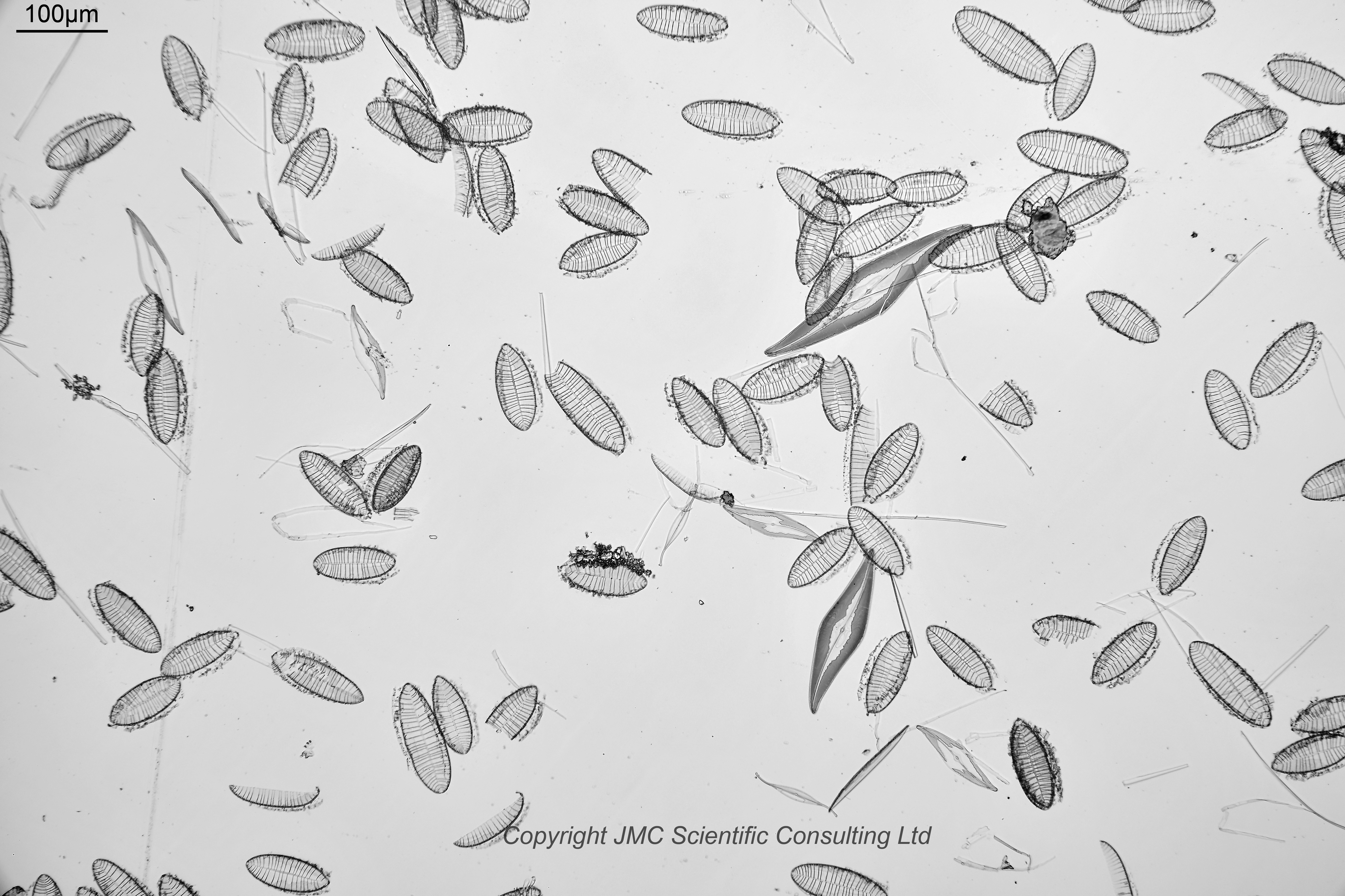
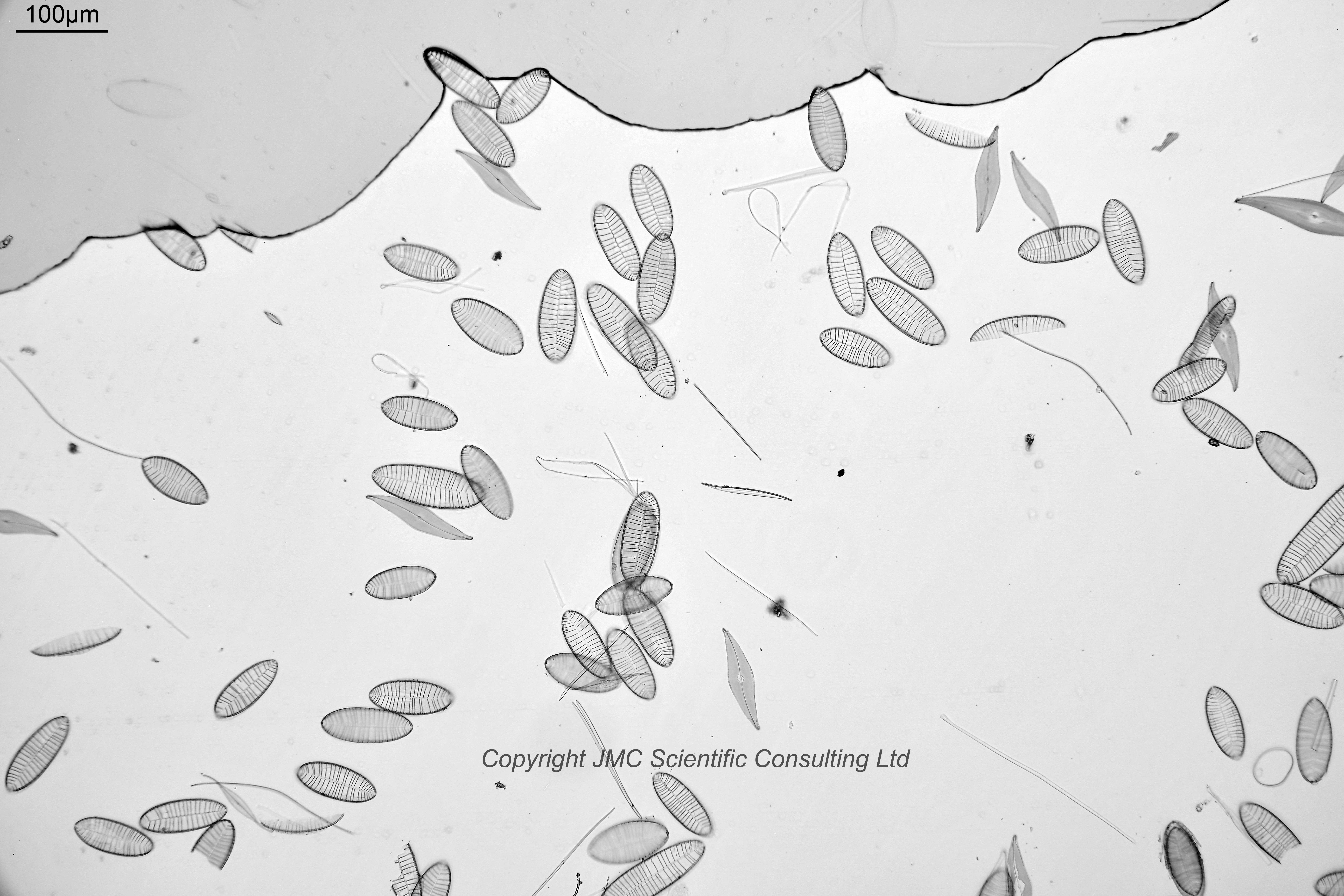
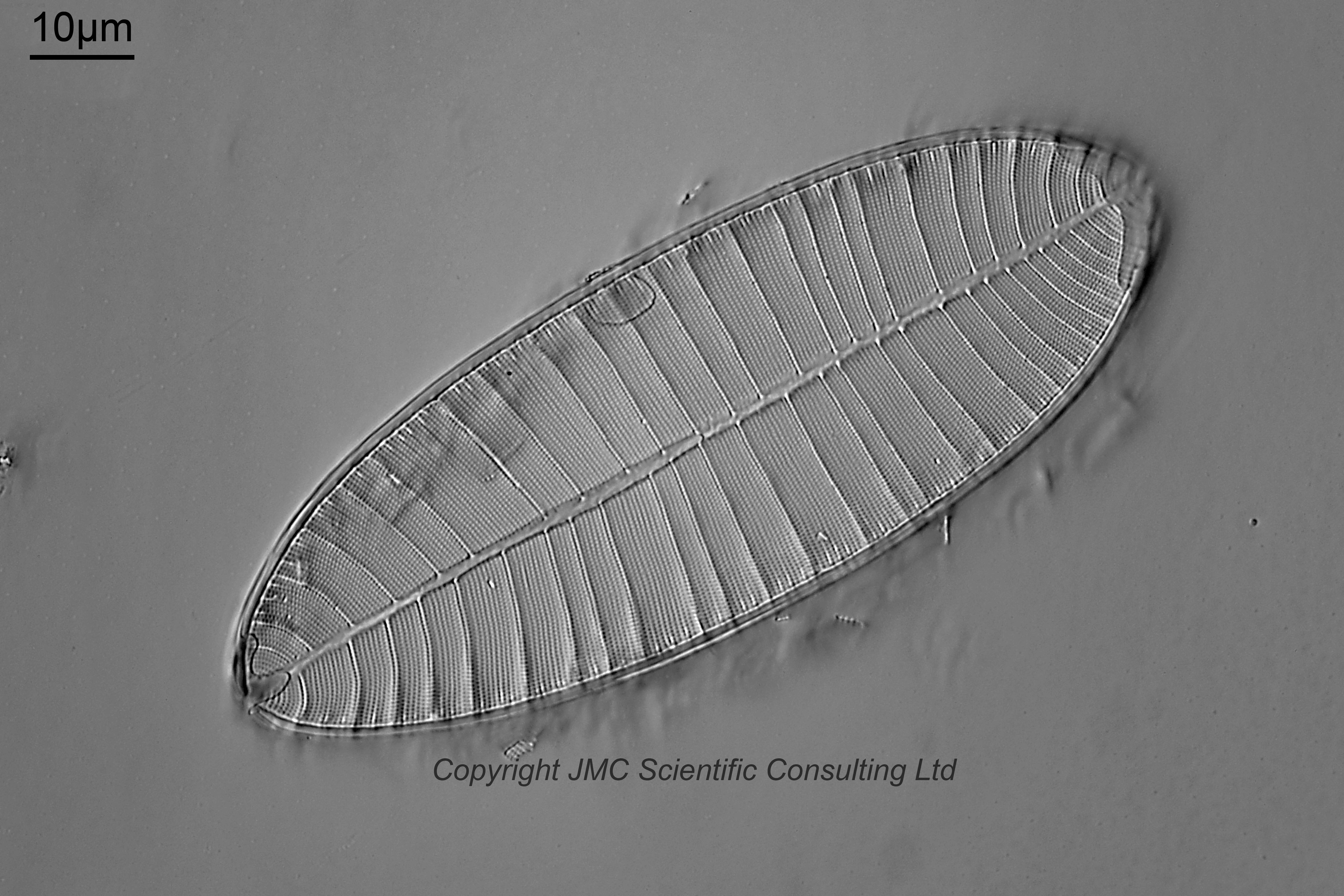
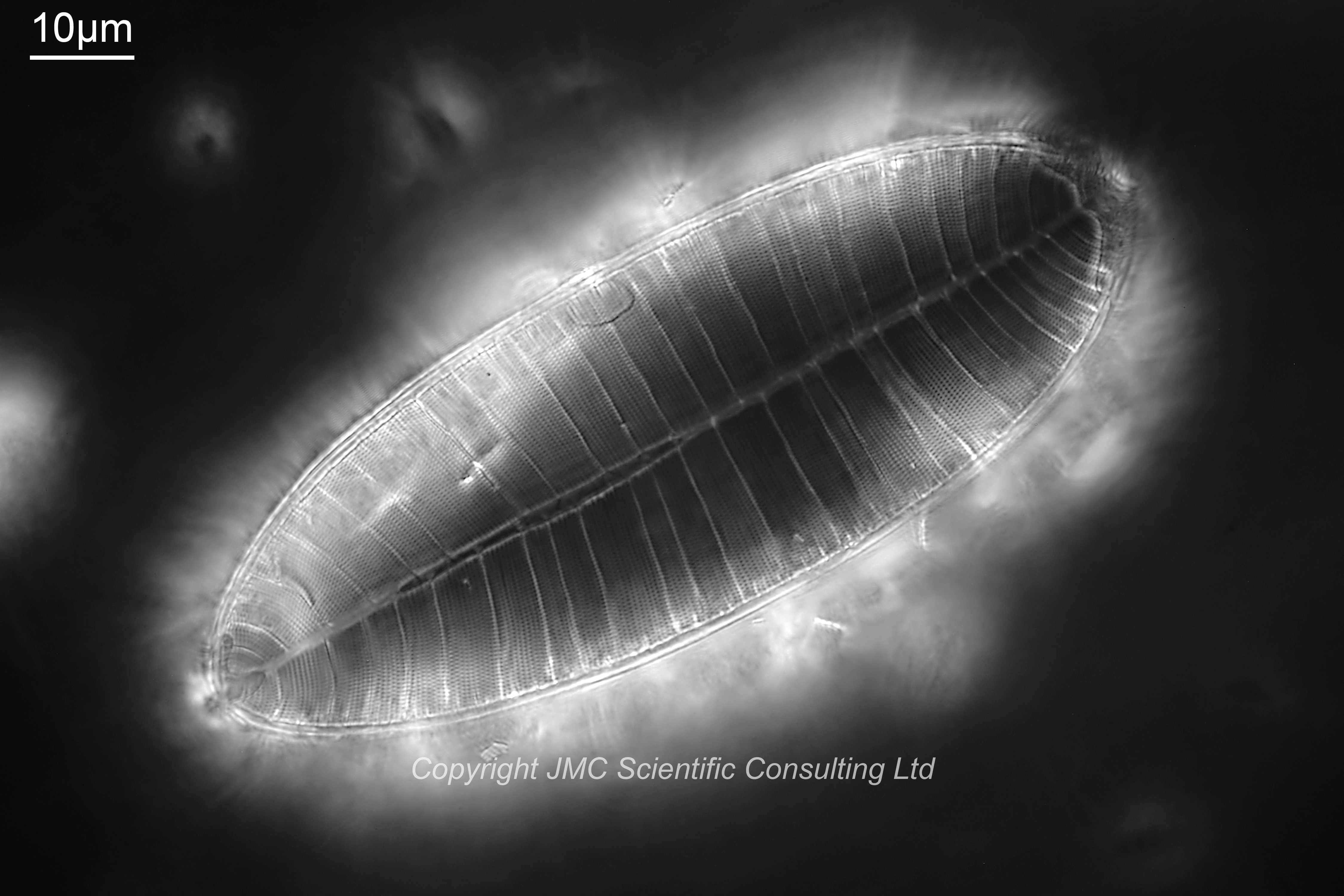
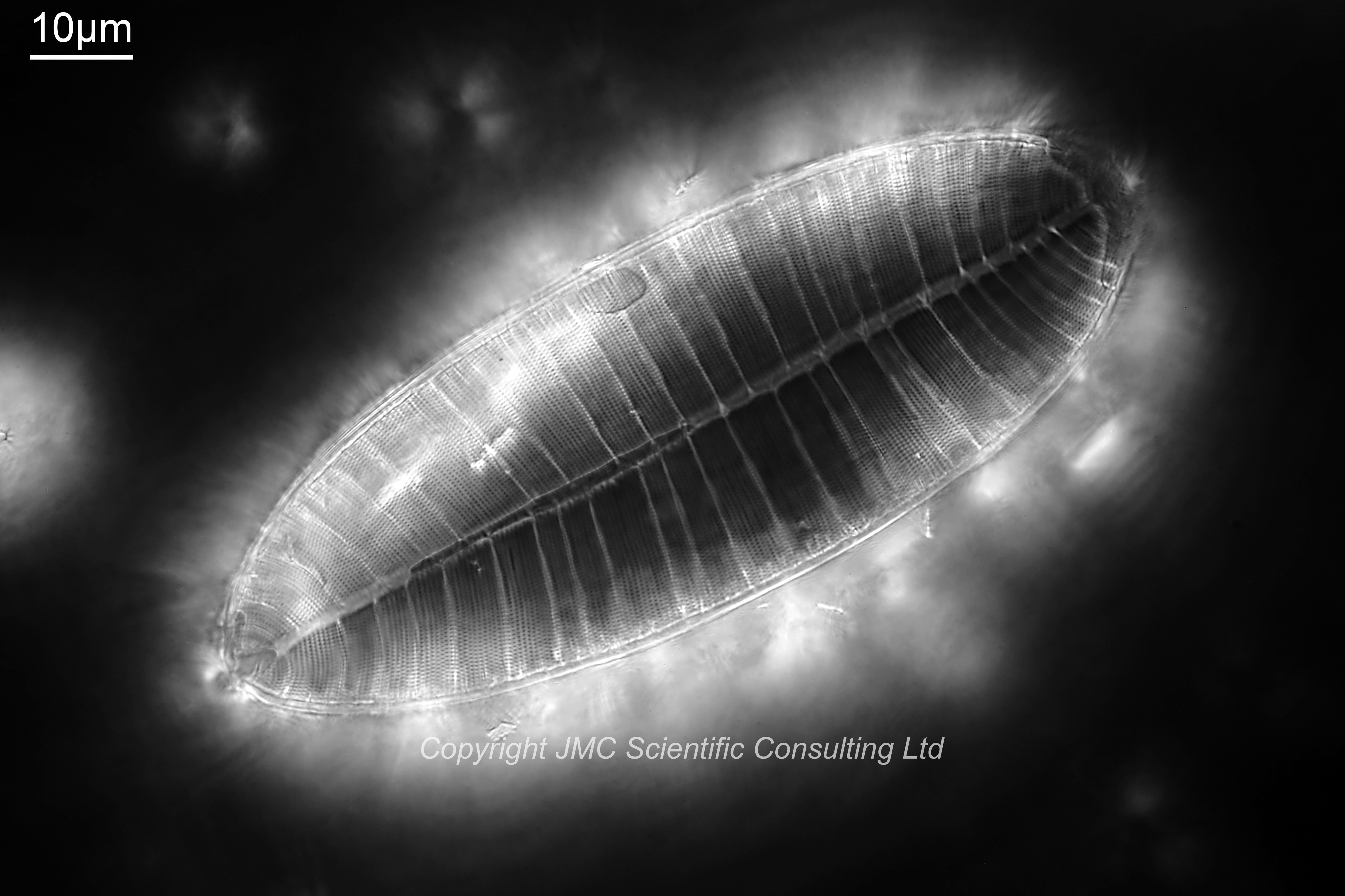
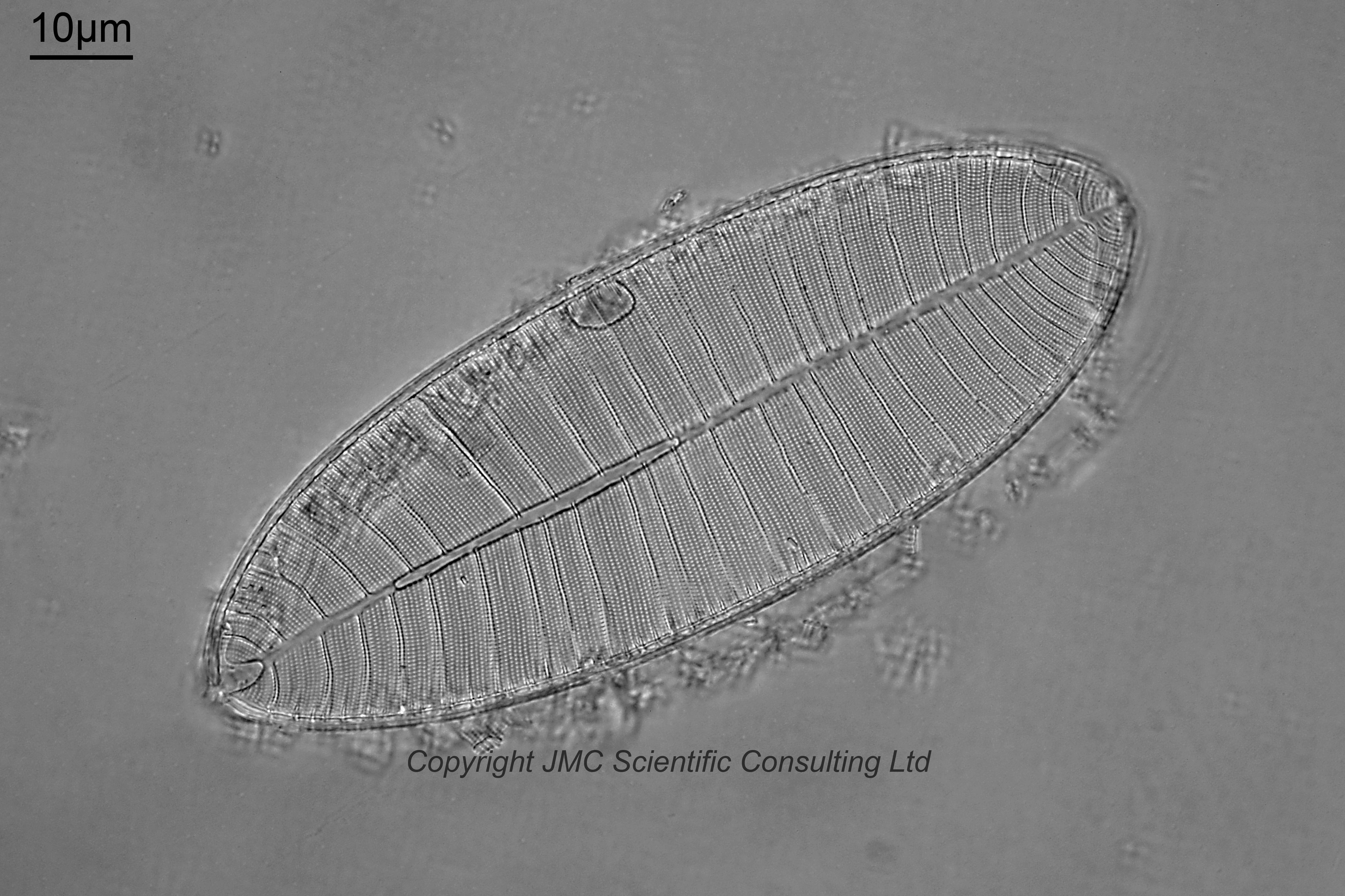
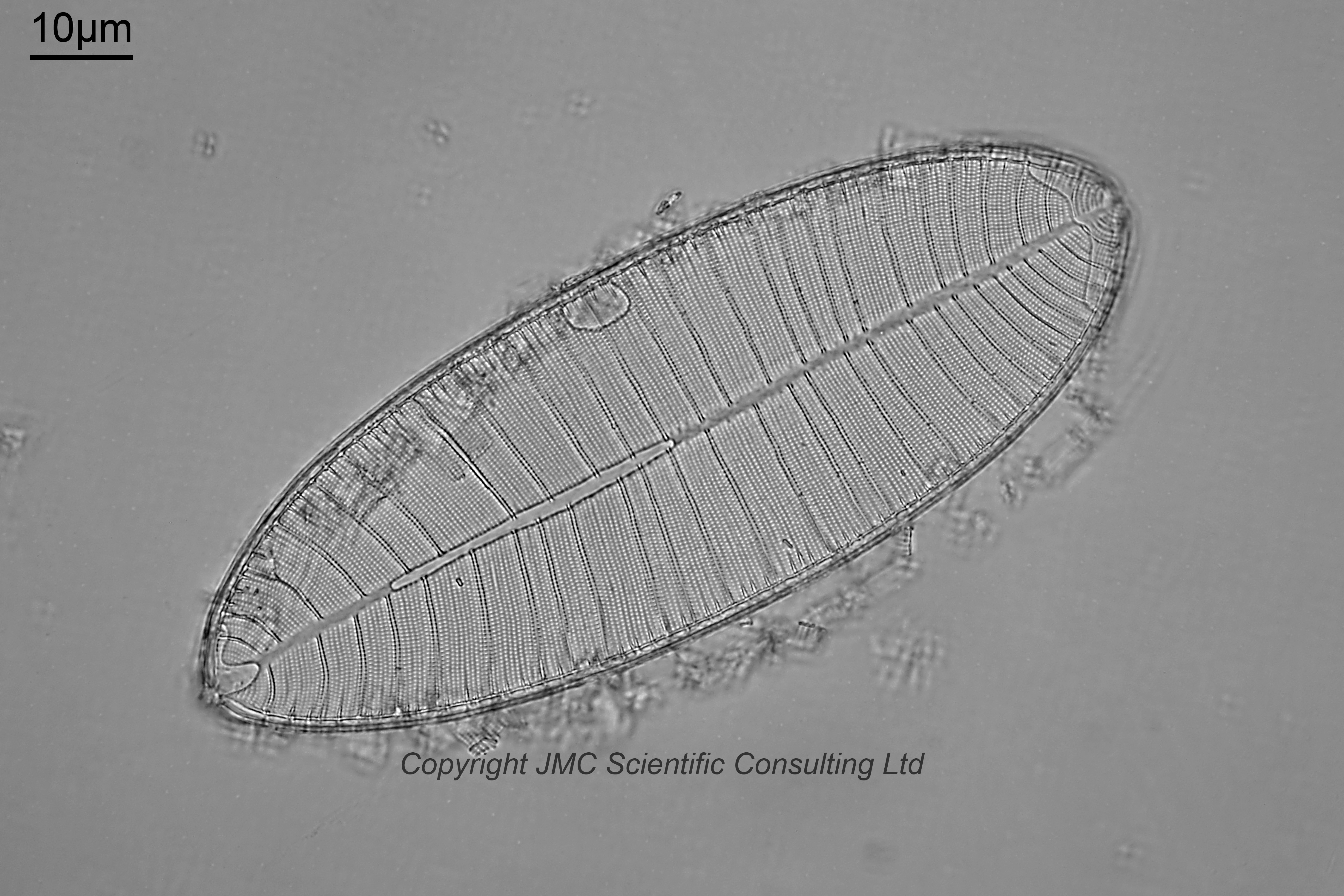
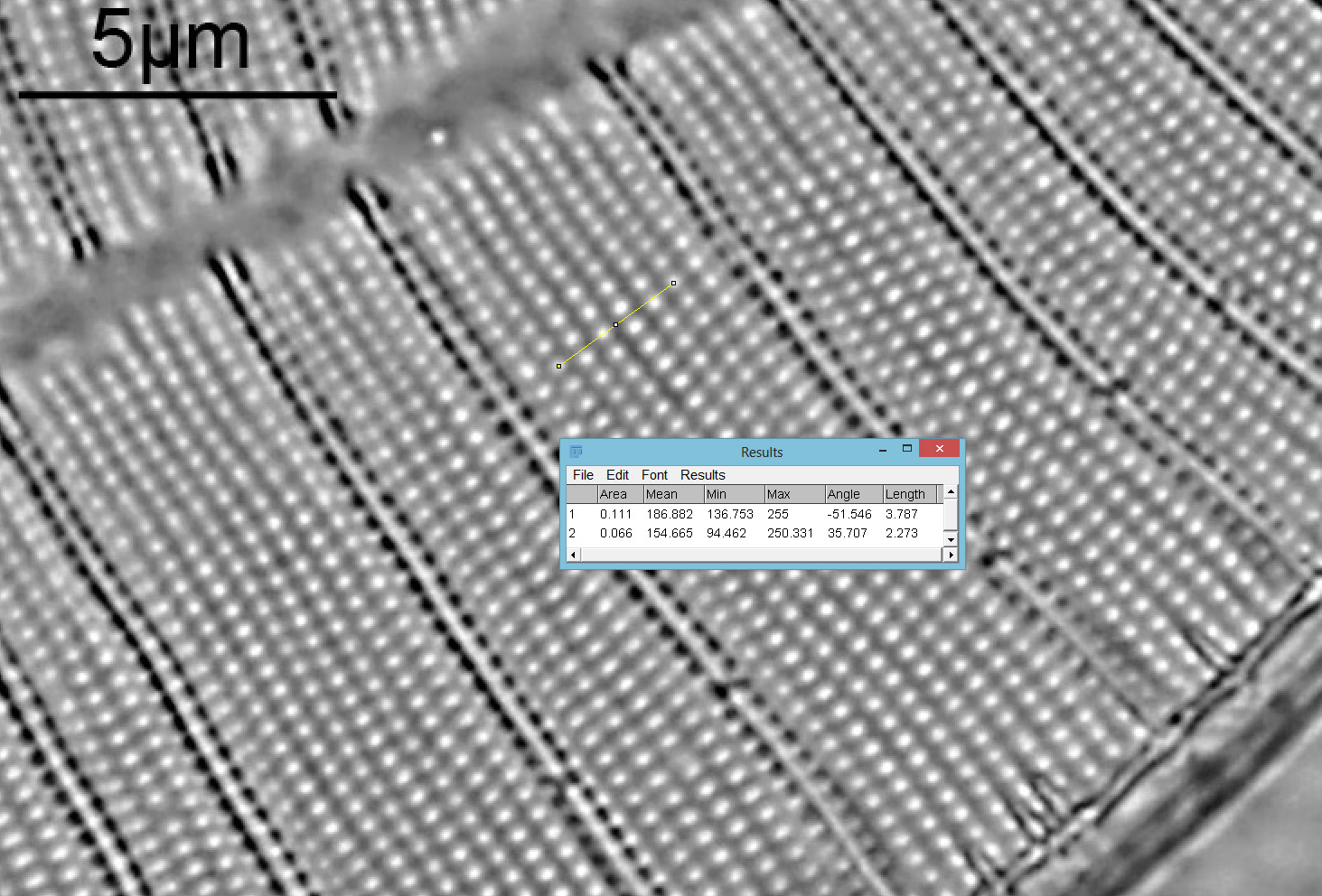
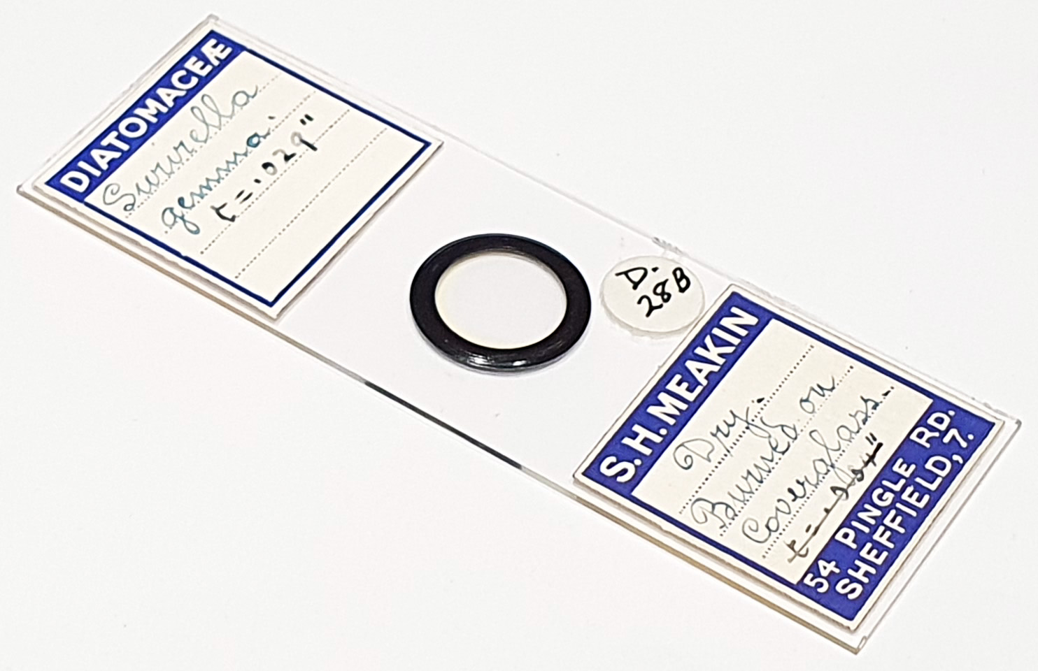
A strew slide of Surirella gemma (and also other species). Dry, burned on coverglass – more on this later. Prepared by Samuel Henry Meakin. Olympus BHB microscope using 450nm or 365nm LED light (details below the images). 63x Leitz Pl Apo 1.4 objective, oil immersion. Olympus Aplanat Achromat condenser, oil immersion, oblique lighting. Reichert Neo 1.18/1.42 dark ground condenser, oil immersion. Olympus Abbe condenser, oil immersion, wide open and closed half way. 2.5x Nikon CF PL photoeyepiece. Monochrome converted Nikon d850 camera. Single images only, no stacking.
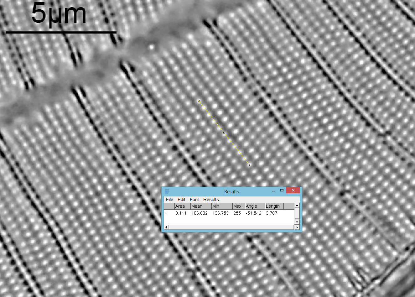
OK, what’s going on here? This is a dry mount where the diatoms have been burned on the coverslip at high temperature. An unusual technique and this is the first slide I’ve seen which specifically states these are burned on as part of the process of making it. This has had the effect of flattening the S. gemma. Normally these are quite curved diatoms, needing multiple images and stacking to get all the puncta in focus. Here they are flat and a single image got the majority of the diatom in focus. There are fragments of material from the the diatom around its perimeter, presumably from this burning, or sintering, process. Oblique lighting witn 450nm gave a nice image, but as this is a dry mount I also wanted to try 365nm light. First though I tried a darkground Reichert Neo condenser with 450nm light, thinking this would give me circular oblique lighting. However it didn’t, it produced a more conventional darkground image. I’ve seen this effect before with dry mounts. Next I tried 365nm with the Reichert dark ground condenser which gave a similar image to 450nm but with slightly better resolution. Next was bright field with an Olympus Abbe condenser and 365nm. I would have liked to have used oblique lighting but the Olympus Aplanat Achromat condenser but that does not let 365nm light through. Condenser wide open there was a bit of flare and reduced contrast, but reducing the iris on the condenser to about half improved this. I took that image and did some image analysis in ImageJ and measured the striae distance as 455nm and the puncta distance as 379nm.