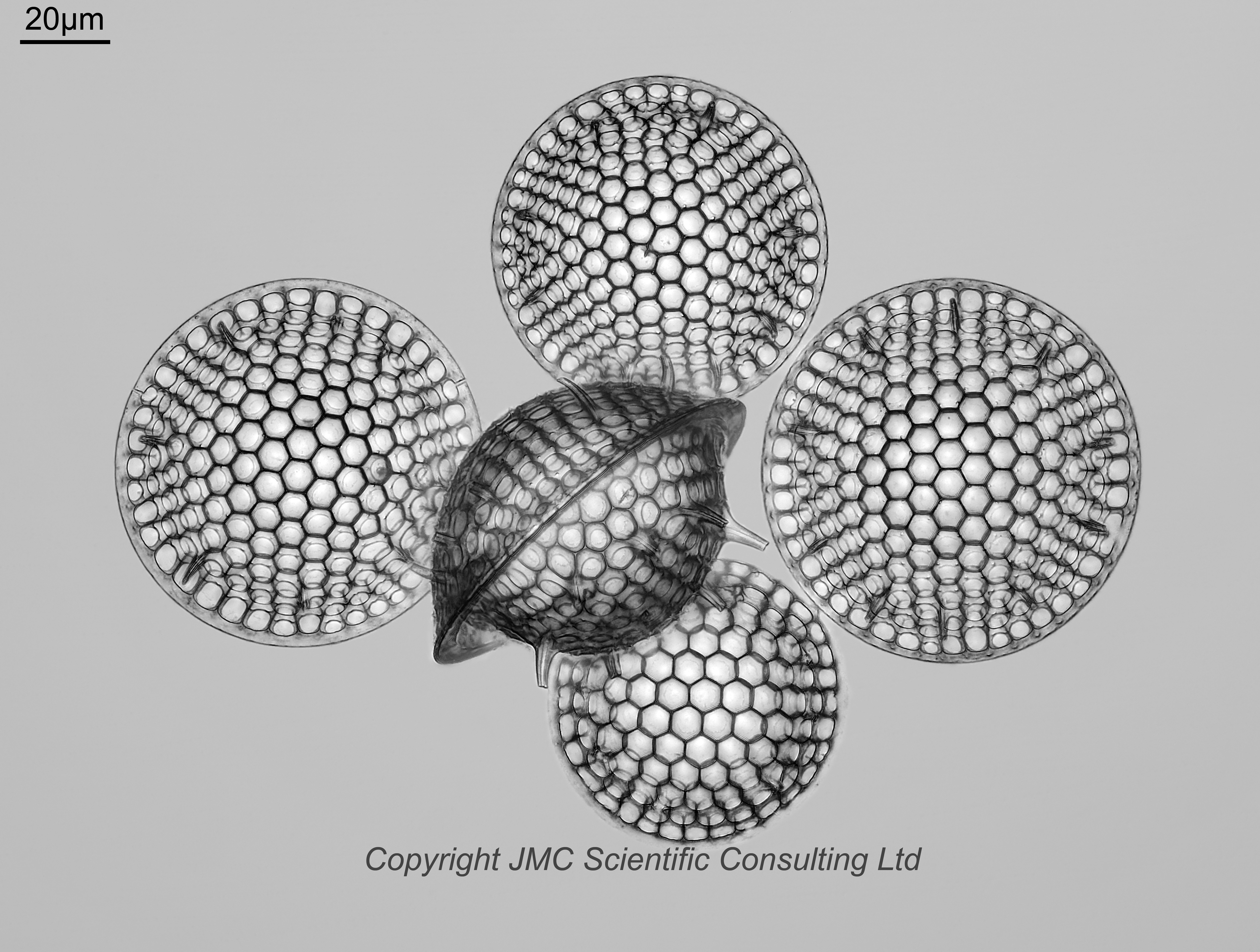
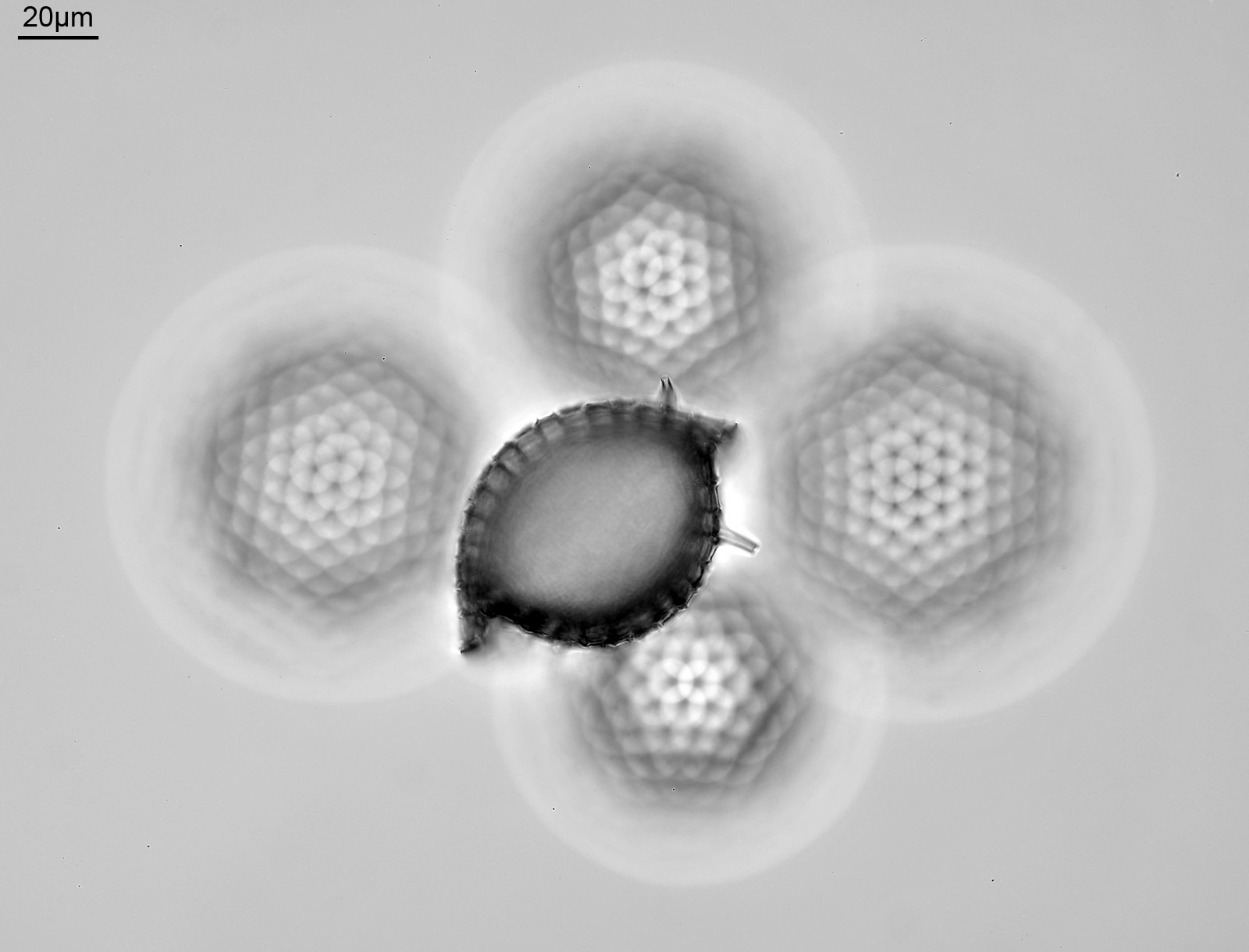
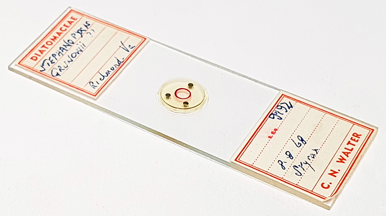
An arrangement of Stephanopyxis grunowii from Richmond, Virginia. Mounted in Styrax. Prepared by CN Walter and dated 8.8.68. The central one is above the other 4 (i.e. closer to the coverslip) and viewed from the side. Olympus BHB microscope using 450nm LED light. 40x Leitz Pl Apo NA 1.00 objective, oil immersion. Olympus Aplanat Achromat condenser, oil immersion, brightfield lighting. 2.5x Nikon CF PL photoeyepiece. Monochrome converted Nikon d850 camera. Challenging to image given the depth of the stack required. The full stack was 181 images (Zerene, Pmax), but I did a second one just of the upper diatom, so that its edges weren’t complicated by the ones below. These were then manually blended together in Zerene. Some really funky looking out of focus highlights for the lower diatoms, when focused above them. Still some artifacts in the final image, but I always seem to struggle with Stephanopyxis.
There are a couple of question marks after the name, so perhaps the mounter wasn’t sure about it. However it does look right to me.