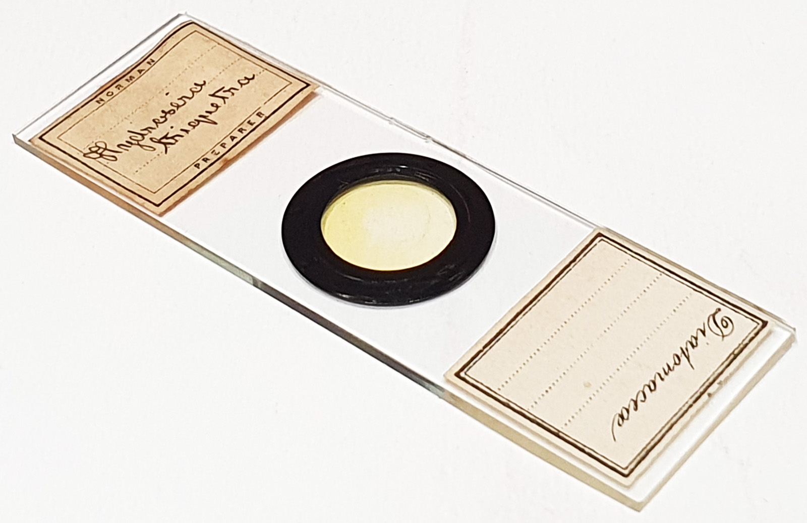
Interesting slide this. A strew slide labelled as Hydrosera triquetra. No location given. Prepared by JT Norman. Olympus BHB microscope using 450nm LED light. As well as the strew, I imaged a couple of diatoms at higher resolution.
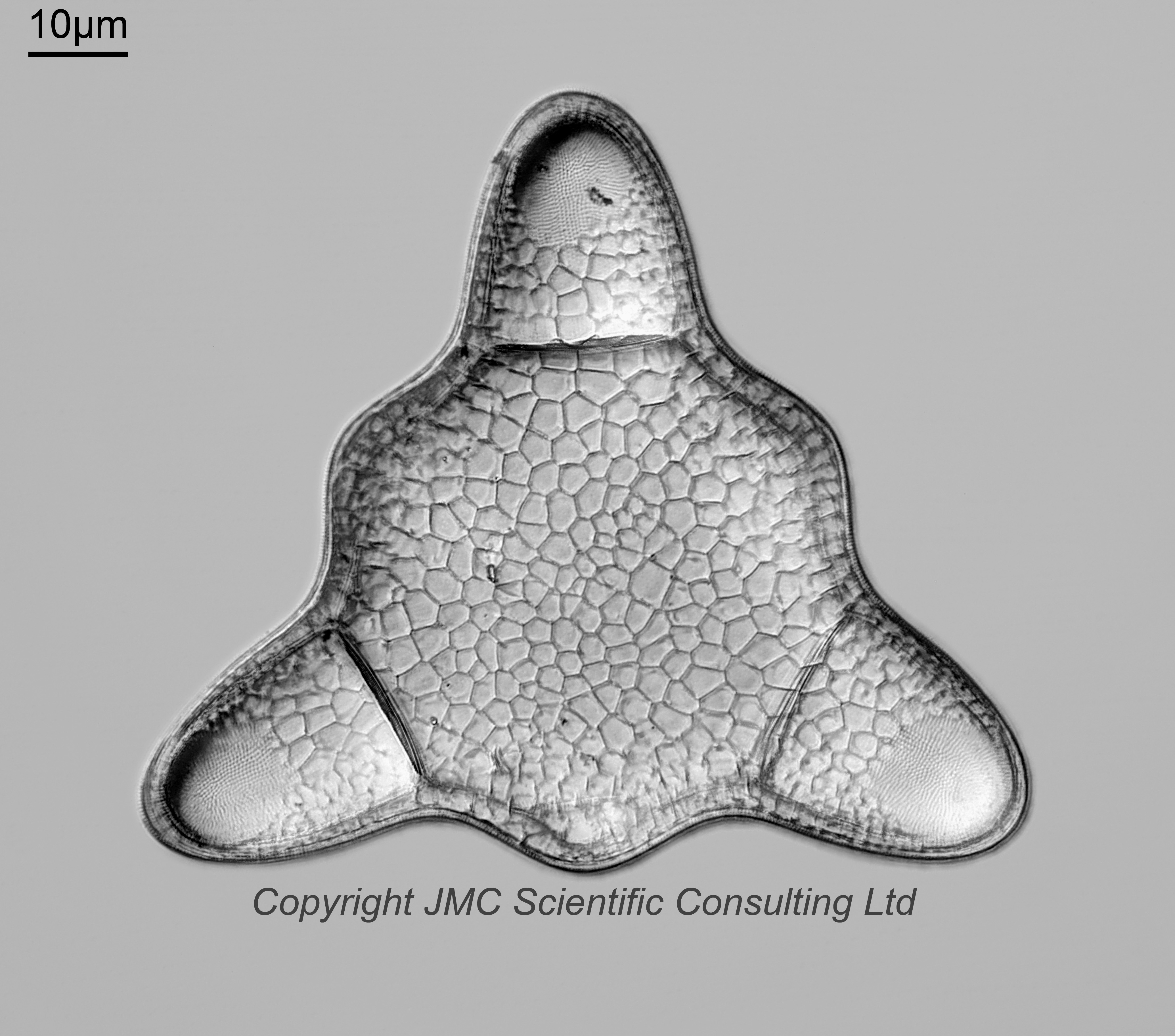
Hydrosera triquetra Wallich, 1858. Published in: Wallich, G.C. (1858). On Triceratium and some allied forms, with figures of the same. Quarterly Journal of Microscopical Science 6: 242-253, pls XII, XIII. Page 251, Plate XIII [13], Figures 1-6. 63x Zeiss Neofluar NA 1.25 objective, oil immersion. Olympus Aplanat Achromat condenser, oil immersion, oblique lighting. 2.5x Nikon CF PL photoeyepiece. Monochrome converted Nikon d850 camera. 27 images stacked in Zerene (Pmax). I ended up using this objective as it has a long working distance, and the usual 40x and 63x Leitz ones I use couldn’t go deep enough.
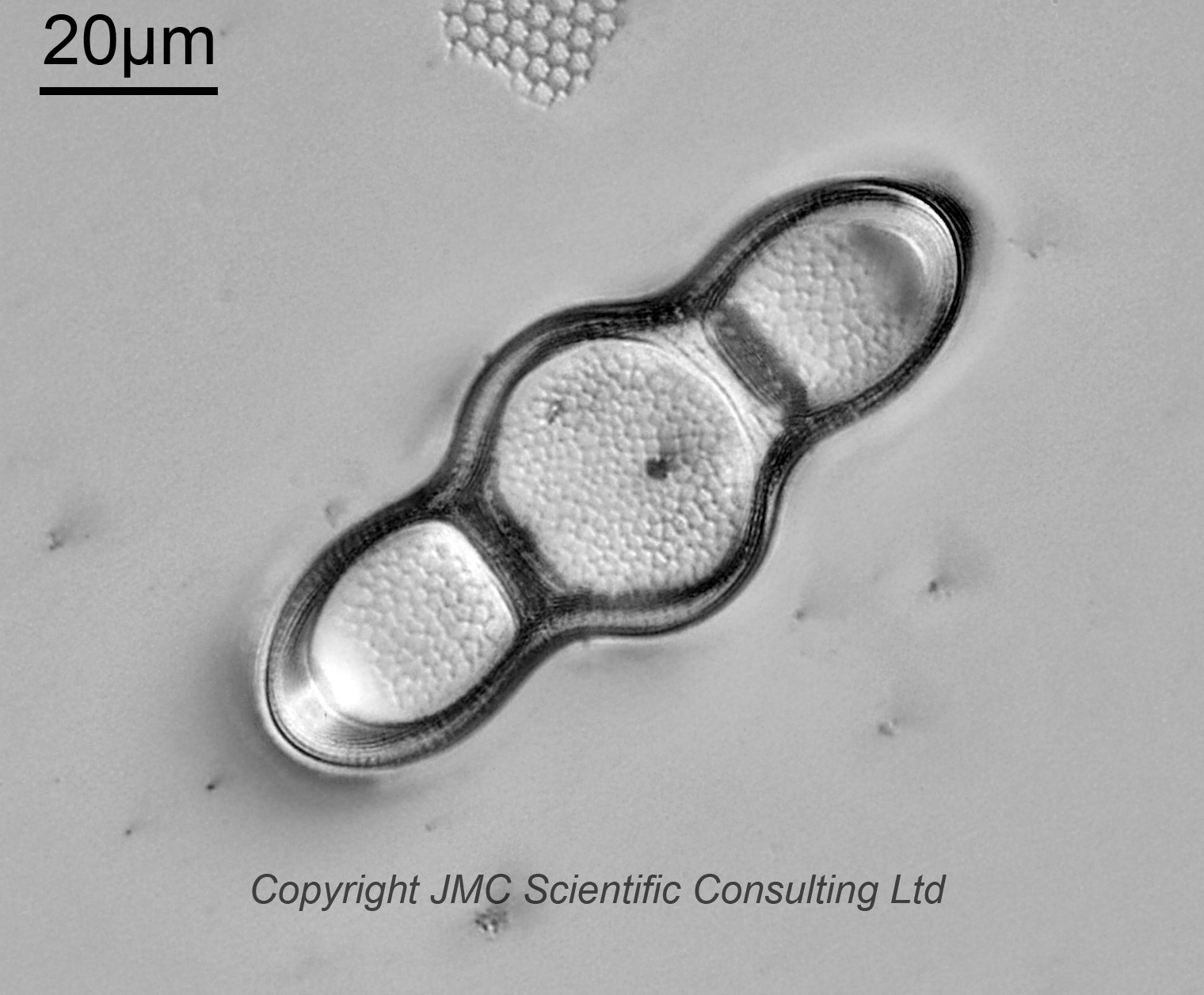
Hydrosera compressa Wallich, 1858. Published in: Wallich, G.C. (1858). On Triceratium and some allied forms, with figures of the same. Quarterly Journal of Microscopical Science 6: 242-253, pls XII, XIII. Page 251, Plate XIII [13], Figures 7-12. I am reasonably sure of this. This one was even deeper than the other one, and I had to just use the 20x Nikon Plan Apo NA objective for this one. 18 images stacked in Zerene (Pmax). An underside view with the rim upwards towards the coverslip.
No location is given on the slide. The diatoms in Wallich, 1858 (which contains H. compressa and H. triquetra) were obtained from the Ganges mangrove forest (the Sundarbands, described as the ‘Gangetic sunderbunds’), in brackish water well within the influence of the tides, and were found growing attached as a soft mossy stratum upon submerged algae or tree stems.