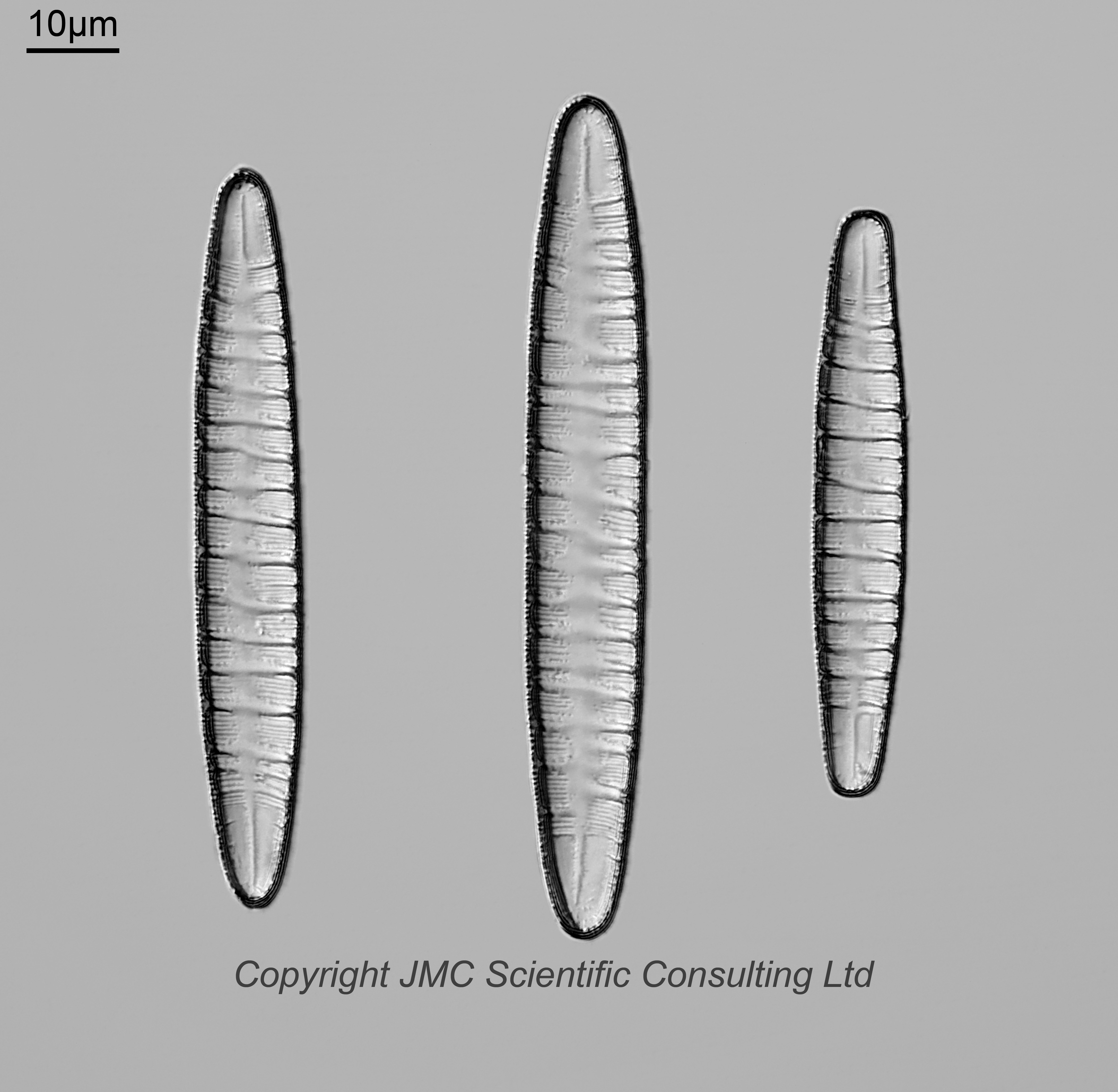
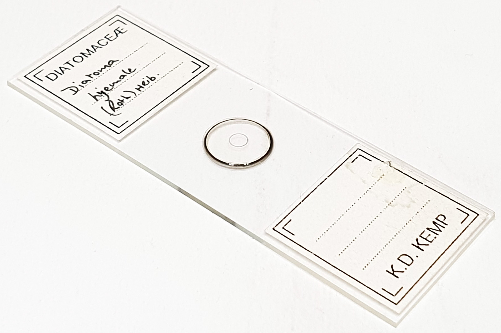
An arrangement of three Diatoma hyemalis (spelt hyemale on the slide). Prepared by Klaus Kemp. Olympus BHB microscope using 450nm LED light. 63x Leitz Pl Apo 1.4 objective, oil immersion. Olympus Aplanat Achromat condenser, oil immersion, oblique lighting. 2.5x Nikon CF PL photoeyepiece. Monochrome converted Nikon d850 camera. 23 images stacked in Zerene (Pmax).
Diatoma hyemalis (Roth) Heiberg, 1863. Published in: Heiberg, P.A.C. (1863). Conspectus criticus diatomacearum danicarum. Kritisk oversigt over de danske Diatomeer. pp. 1-135, 6 pls. Kjøbenhavn: Wilhelm Priors Forlag. Page 58. This name is currently regarded as a synonym of Odontidium hyemale (Roth) Kützing, 1844. Published in: Kützing, F.T. (1844). Die Kieselschaligen Bacillarien oder Diatomeen. pp. [i-vii], [1]-152, pls 1-30. Nordhausen: zu finden bei W. Köhne. Page 44, Plate 17, Figure 4.
Sub-apical rimoportulae most visible on the left hand one towards the bottom, and the right hand one near the top, and (less obvious) nearer the center of the middle one.