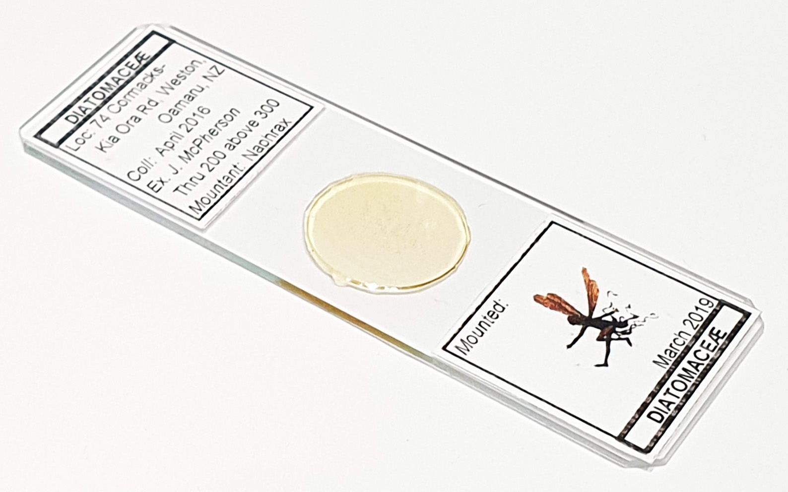
This is going to be a long page as I am including images from 4 slides. These contain material from Cormacks (location 74 Cormacks, Kia-Ora Road, Weston, Oamaru, New Zealand). The material was collected in April 2016 by J. McPherson. It is mounted in Naphrax and the slides were prepared by Little Imp (Mike Samworth and Steve Gill). The set of 4 slides has material which has gone through different stages of filtering – above 72 Mesh, 72-120 Mesh, 120-200 Mesh and 200-300 Mesh. I’ll show each slide as a wide field of view as an example of what is present, along with some higher magnification images. All images were done using an Olympus BHB microscope and 450nm LED light. For the high magnification images, a 63x Leitz Pl Apo NA 1.40 objective with oil immersion was used, unless noted. Olympus Aplanat Achromat condenser, oil immersion, with a mixture of brightfield and oblique lighting. 2.5x Nikon CF PL photoeyepiece. Monochrome converted Nikon d850 camera. Stacking done using Zerene (Pmax). I’ve made an attempt at naming them using various sources including the Oamaru Diatoms website, Schmidt’s Atlas, and the book ‘Oamaru Diatoms’ by Desikachary and Sreelatha. However there are some discrepancies between images from different sources, so in cases where I am less than certain I have included question marks after the names in the images. Enough chat, on with the images.
Above 72 mesh slide
This slide had the largest material present, as it was filtered through the largest mesh. Lost of sponge spicules as well as diatoms.
The image I chose from this slide was that of a ‘Lithisted dermal spicule of Discodermia‘. Not a diatom, but I am including here as it was the only one I found on this slide. This was imaged using a 40x Leitz Pl Apo NA 1.00 objective with oil immersion, and brightfield lighting.
Discodermia is apparently a deep sea sponge. Examples of this came from a couple of locations. For example, Päule Heck’s book “Die fossilen Diatomeen Oamarus (Neuseeland)” shows ‘Lithisted dermal spicule of Discodermia‘ on page 189. Also, Plate XIV, Fig 11, shows ‘Lithistid dermal spicule of Discodermia‘. “On the Sponge-remains in the Lower Tertiary Strata near Oamaru, Otago, New Zealand.”, G. Jennings Hinde Ph.D., and W. Murton Holmes. The Journal of the Linnean Society of London. Zoology. 1892, 24(151), pp. 177-262, Plates VII-XV. Note slightly different spellings.
72-120 mesh slide
This next slide in the series, filtered through slightly smaller mesh. Still lots of sponge spicules in addition to diatoms.
For the Hercotheca mammillaris image (also spelt Herotheca mamillaris) this is quite a tentative ID based primarily on Desichary and Sreelatha’s book, Plate 142, Figures 13 and 14. Imaged using oblique lighting.
The ‘Discoidal spicule of Erylus’ is a tentative ID based on Plate XIV, Fig 33 in “On the Sponge-remains in the Lower Tertiary Strata near Oamaru, Otago, New Zealand.”, G. Jennings Hinde Ph.D., and W. Murton Holmes. The Journal of the Linnean Society of London. Zoology. 1892, 24(151), pp. 177-262, Plates VII-XV. Again, not a diatom, but an interesting part of the makeup of the material from this site. Imaged using brightfield lighting.
120-200 mesh slide
Filtered through a smaller mesh size again, starting to get much less sponge material and a higher proportion of diatoms in the mix with many either intact or almost intact.
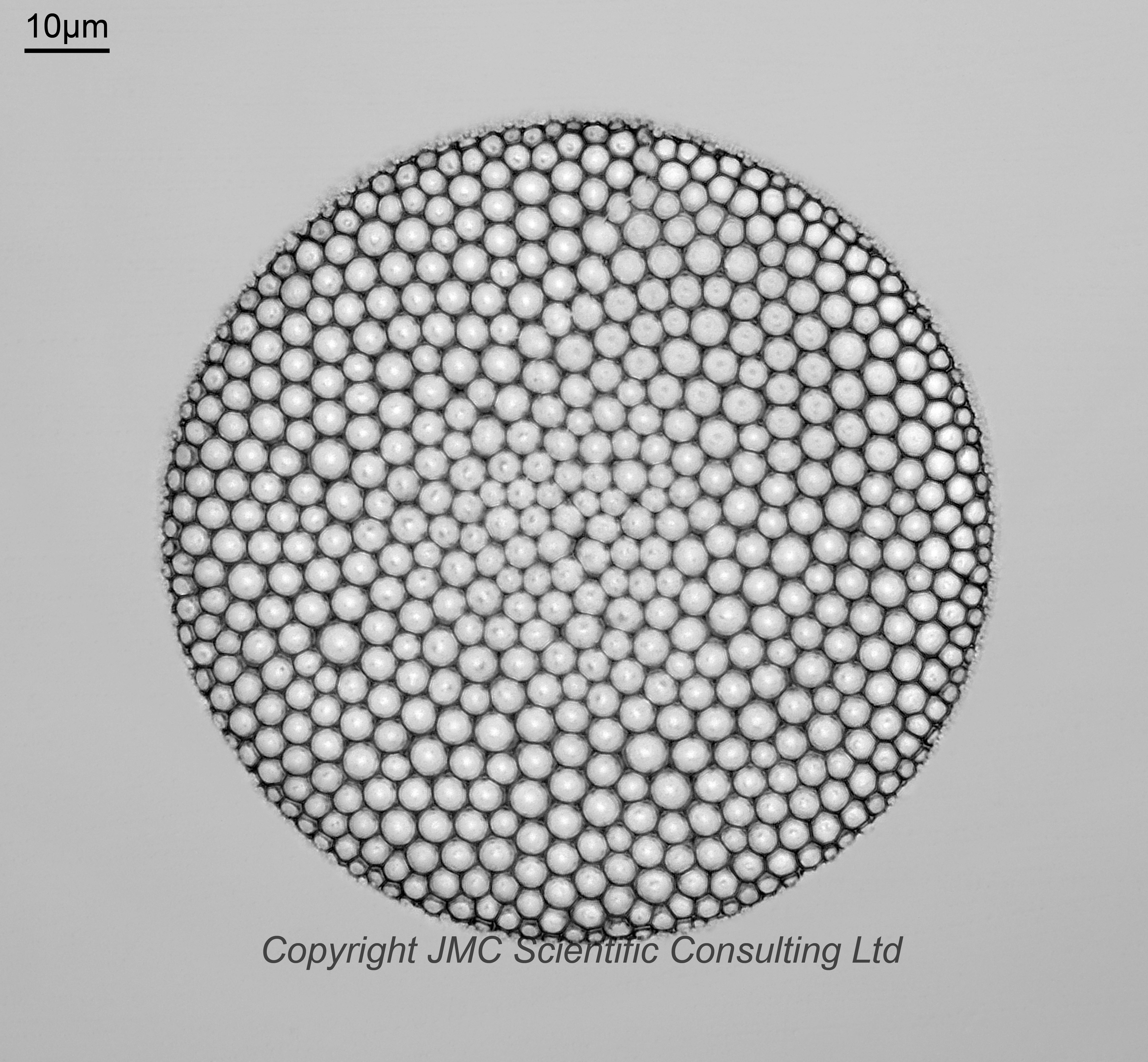
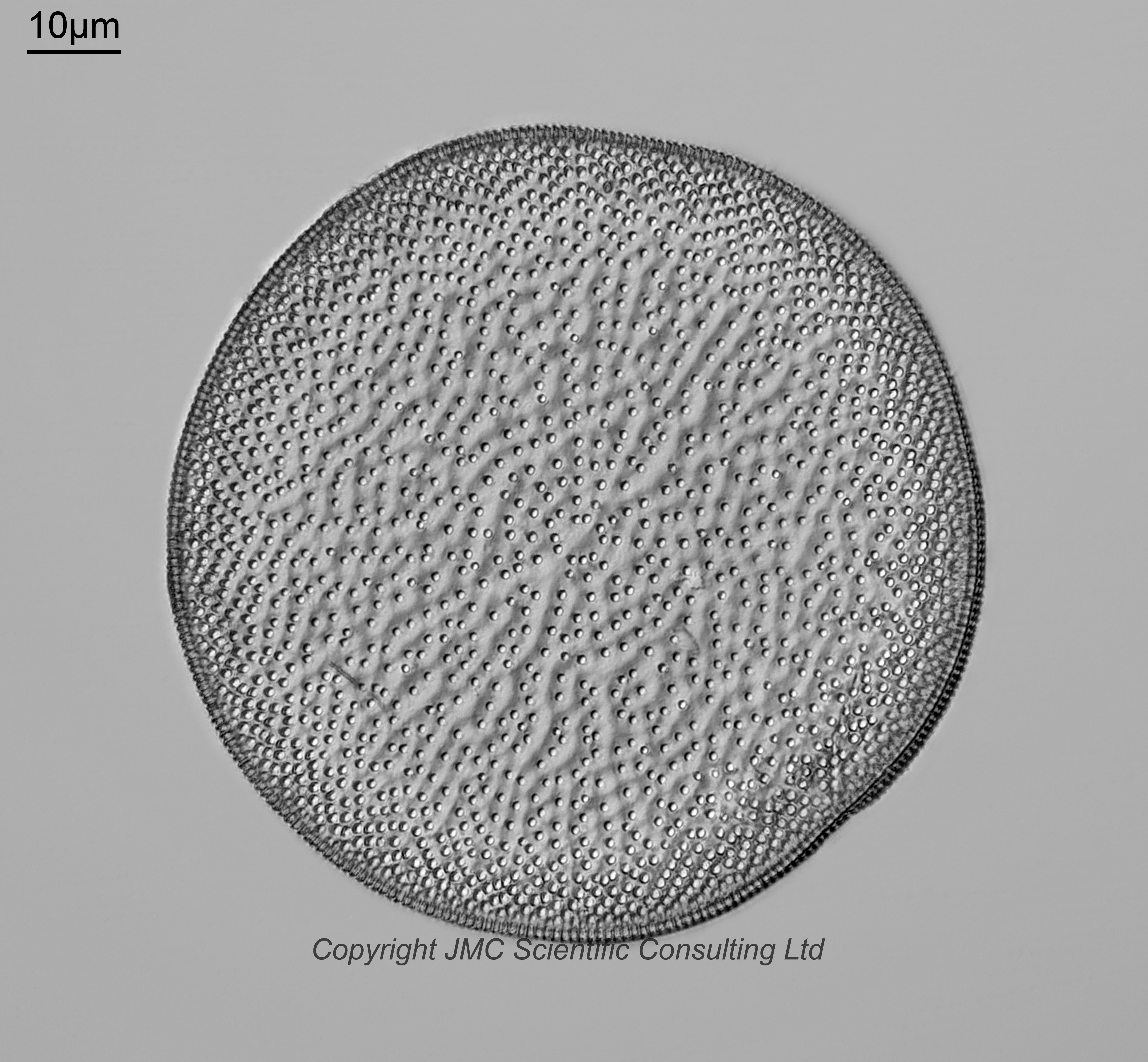
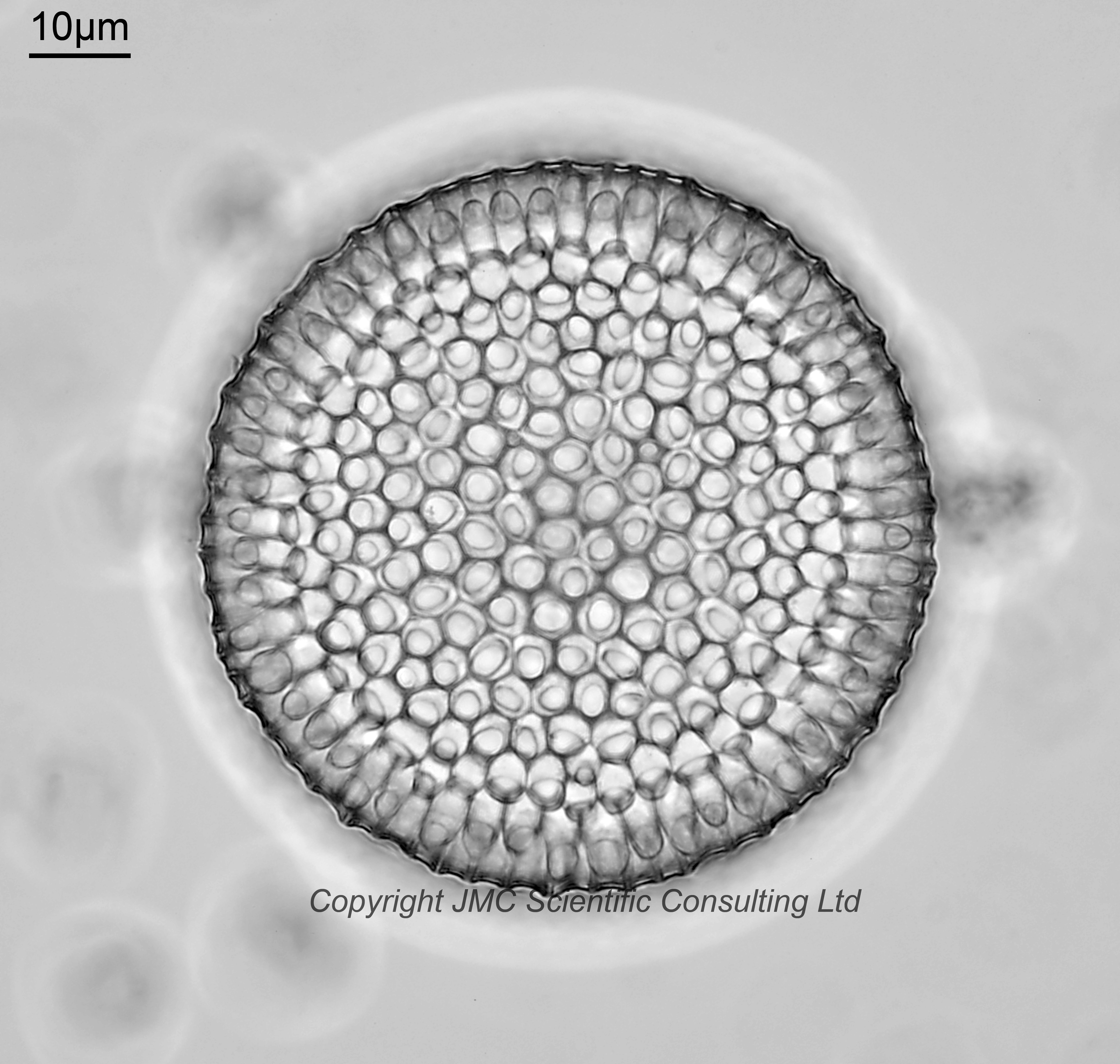
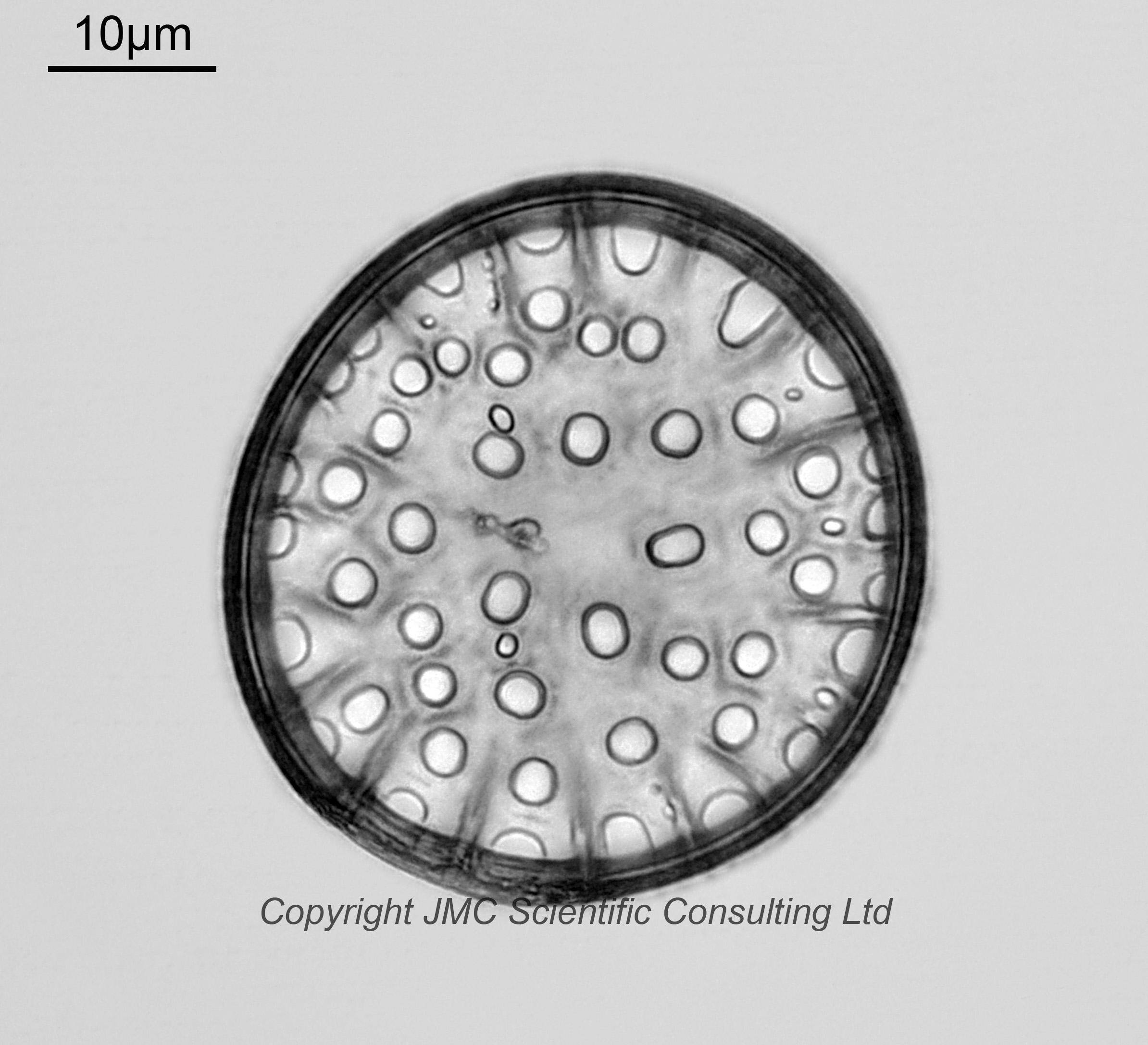
Coscinodiscus decrescens. I’m not sure about this one and it is a tentative ID, as it could be another Coscinodiscus or Azpeitia species. Imaged using brightfield lighting.
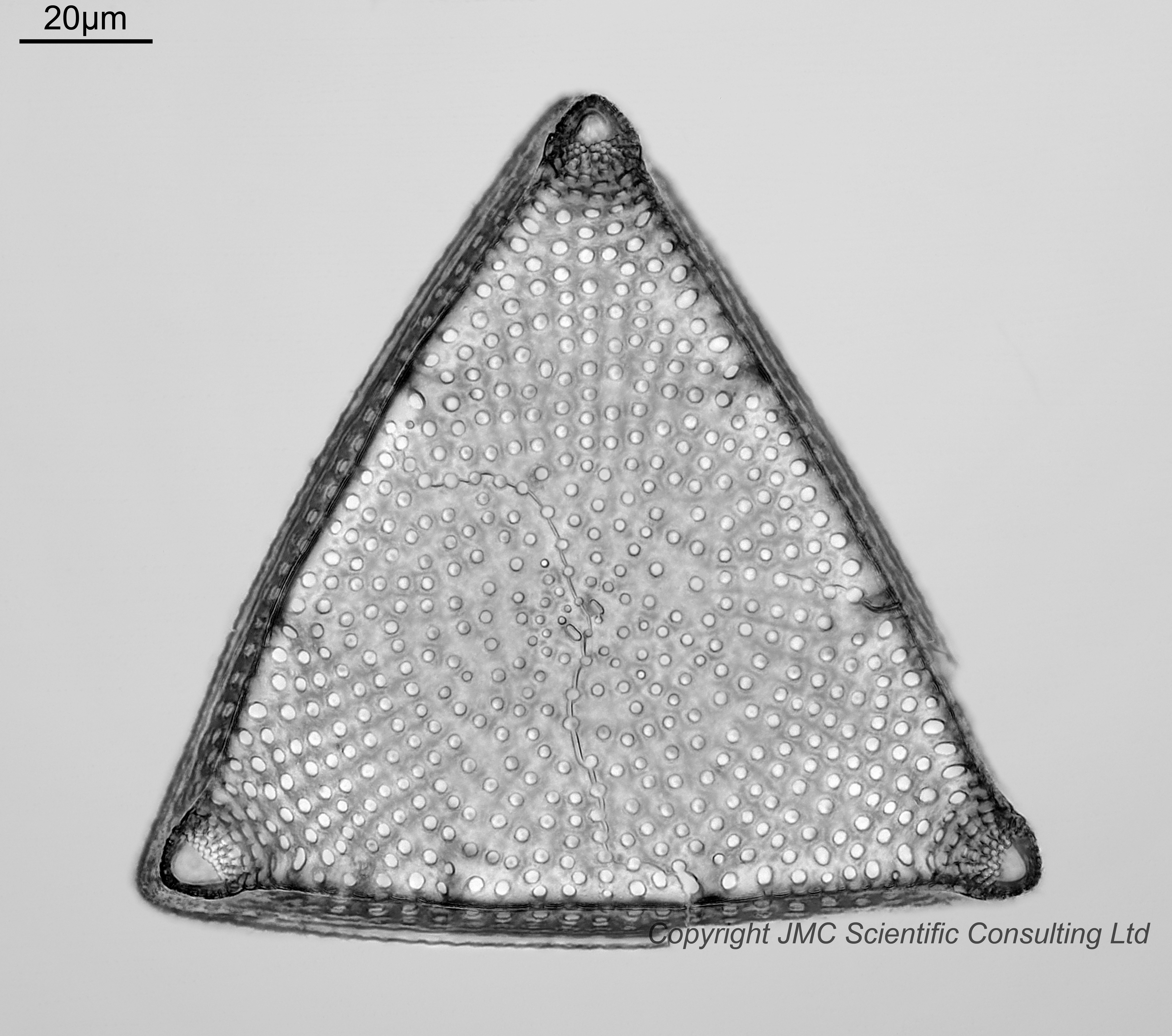
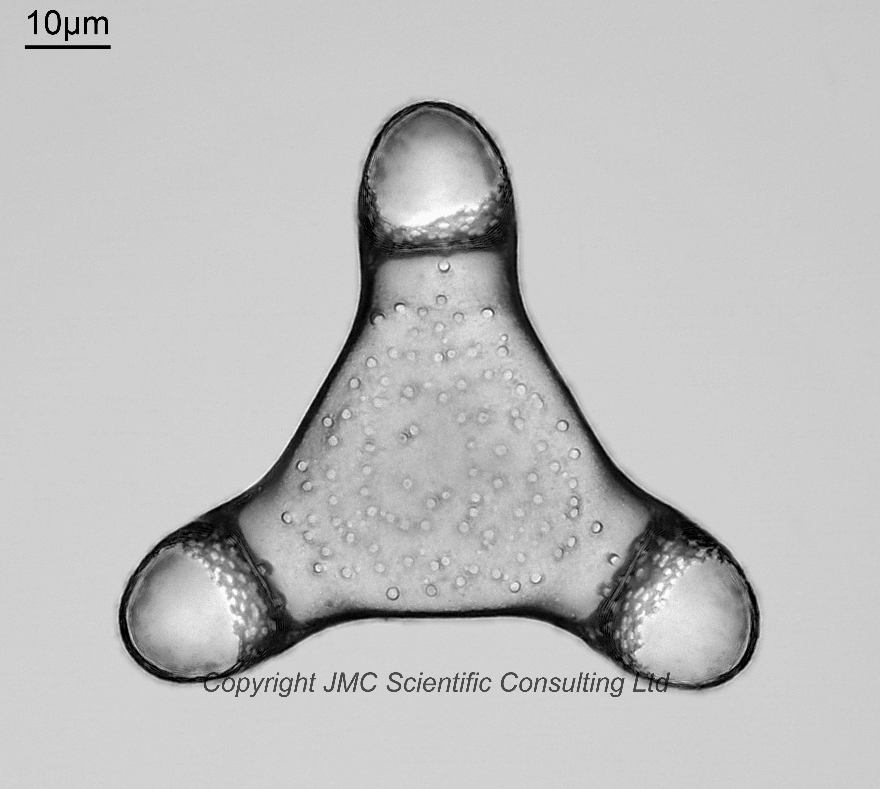
Entogoniopsis pseudonervata. This I am confident about. Old name Trigonium pseudonervatum. Two examples, one viewed from below one from above. Imaged using brightfield lighting.
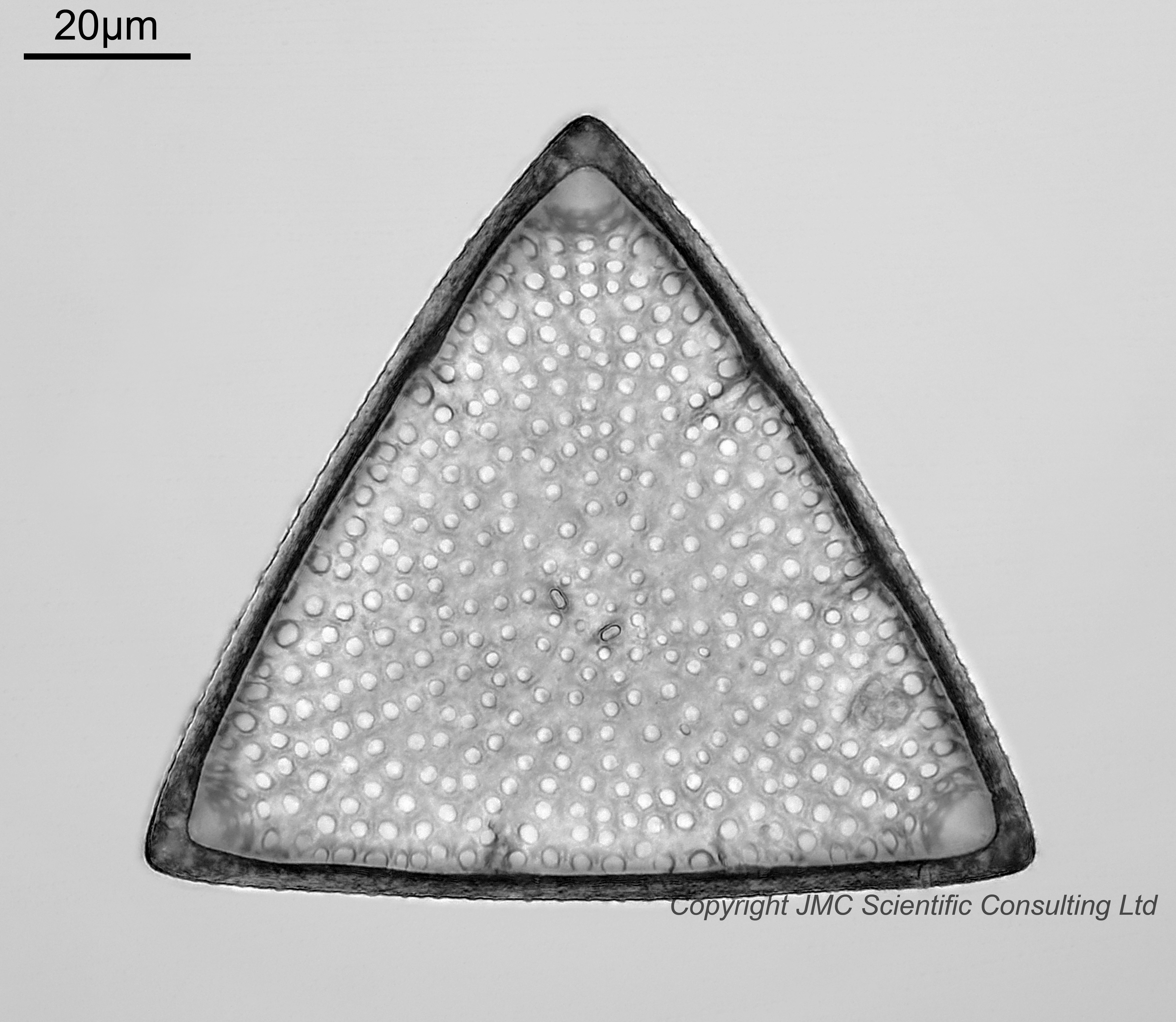
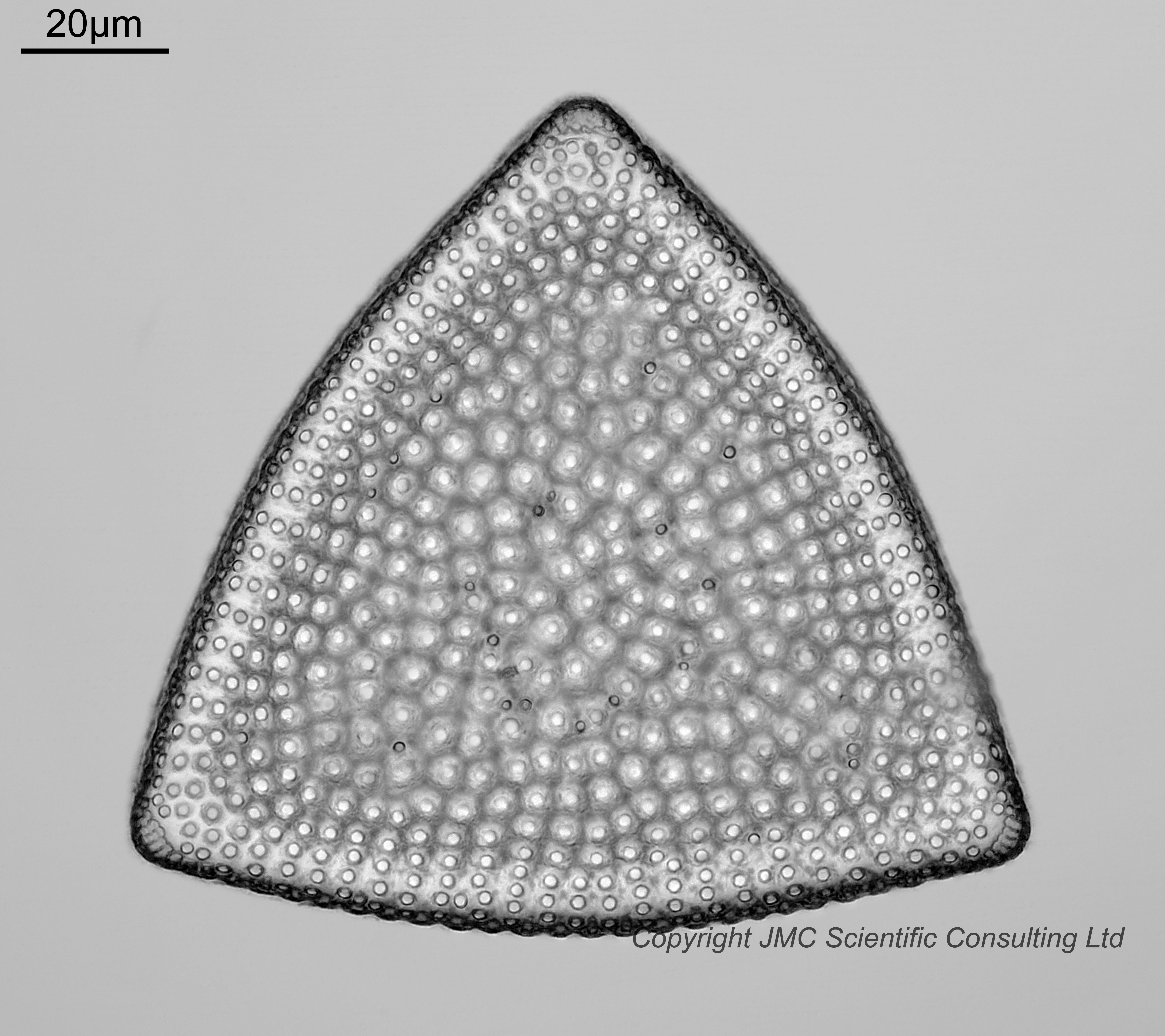
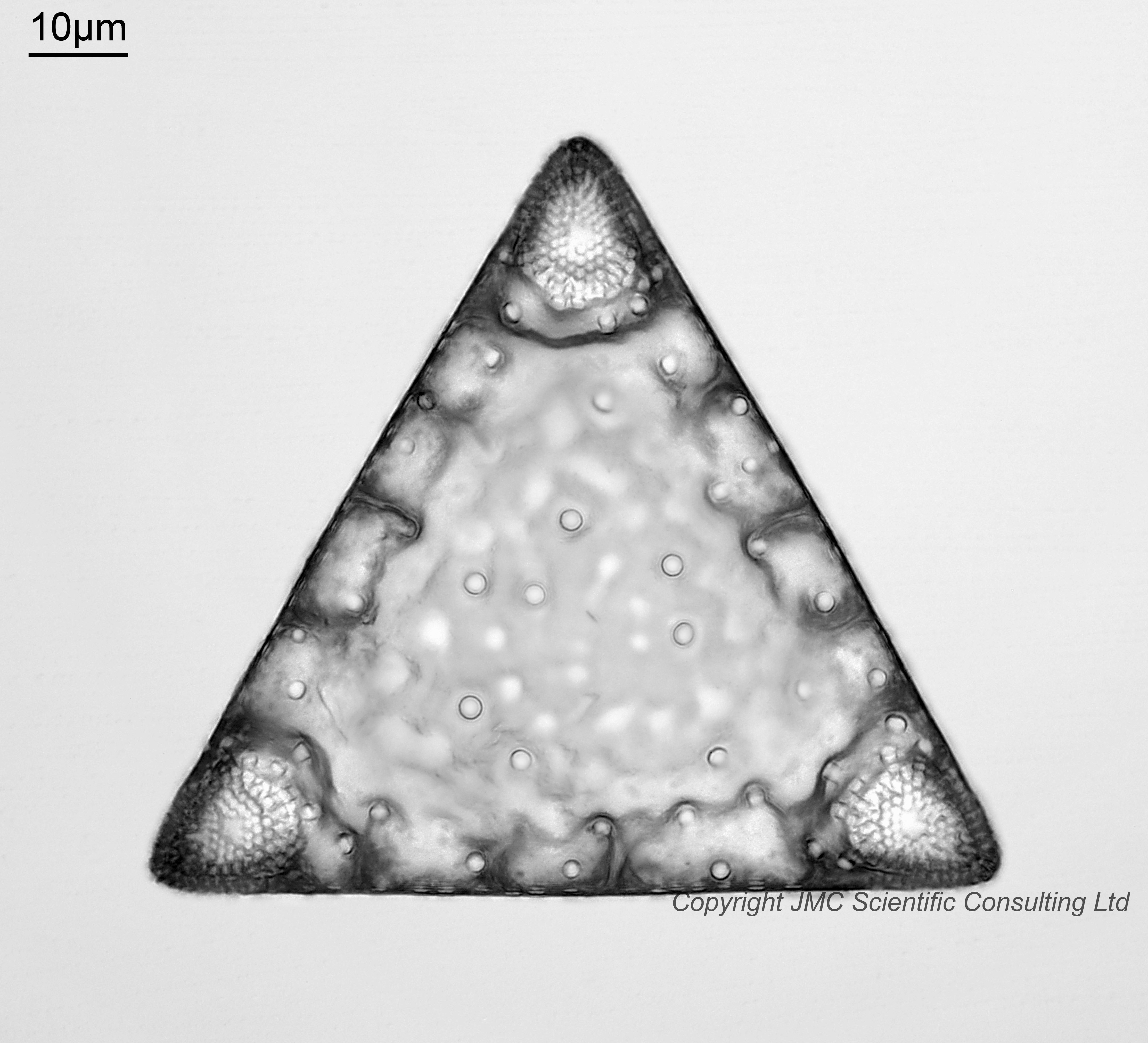
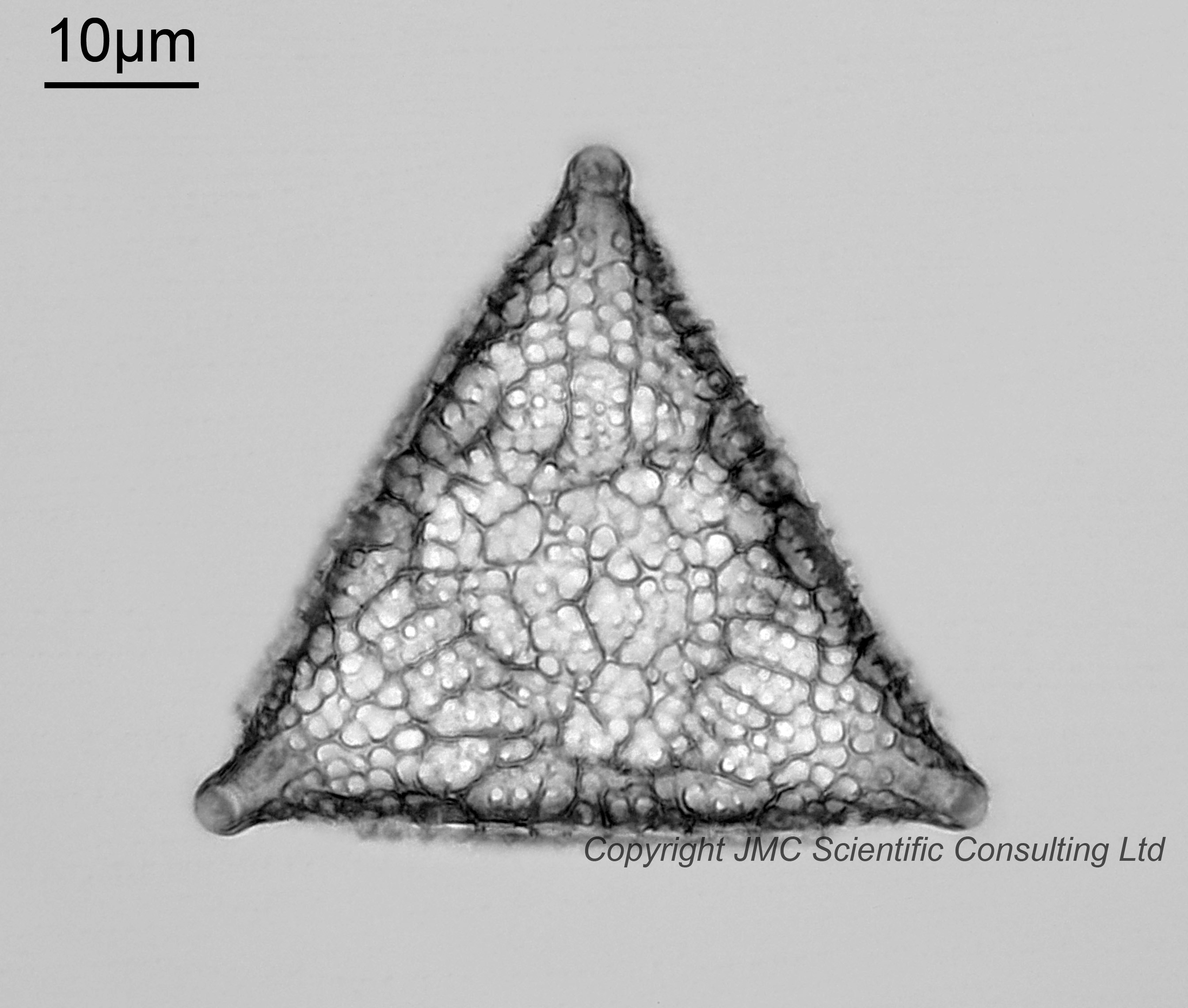
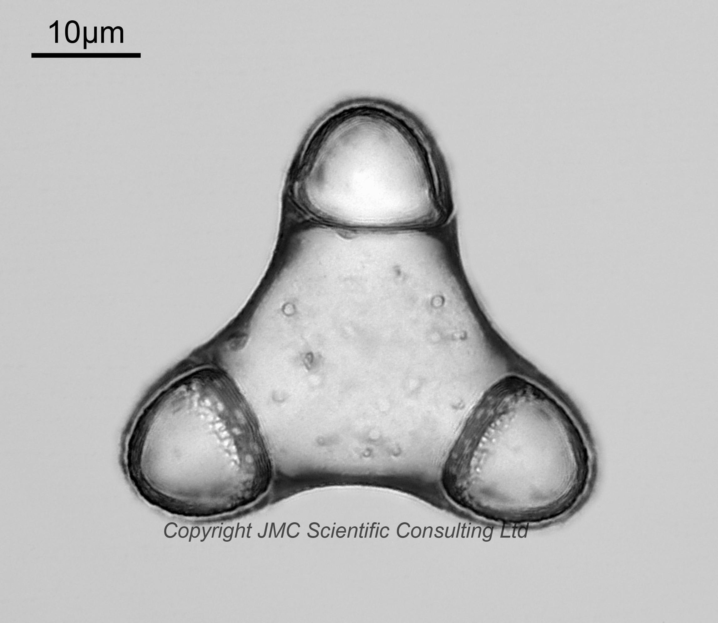
Entogoniopsis major. Old name Triceratium majus. Viewed of the underside and imaged using brightfield lighting.
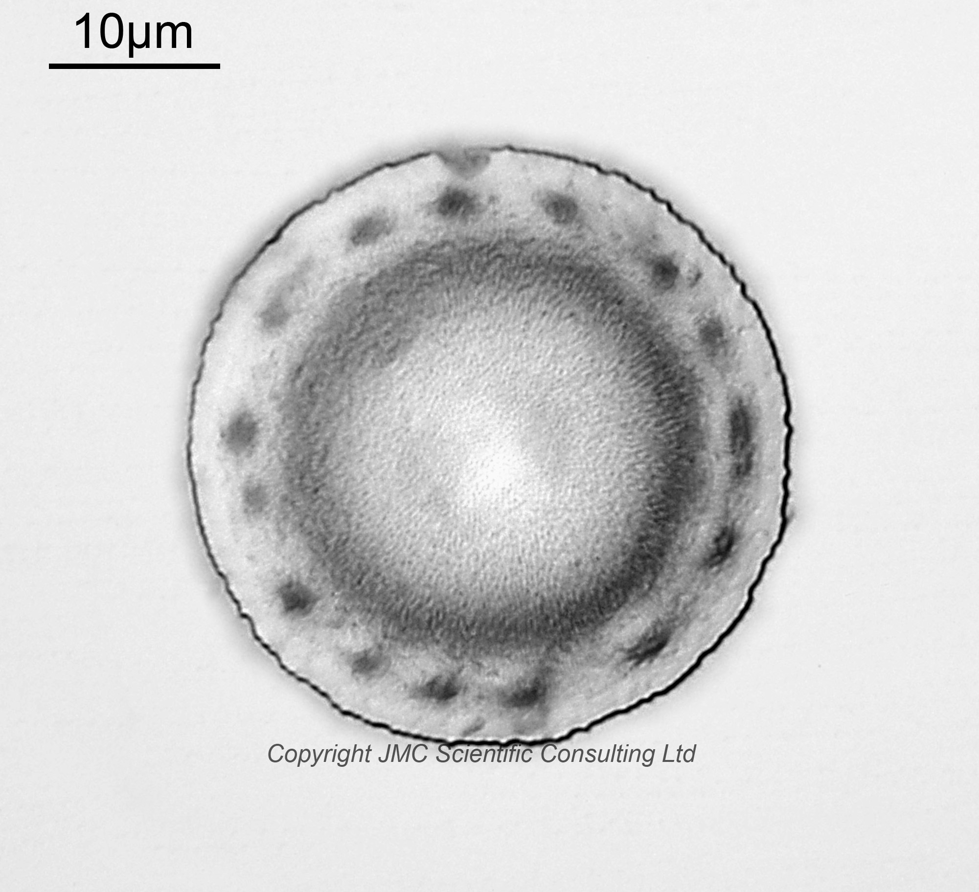
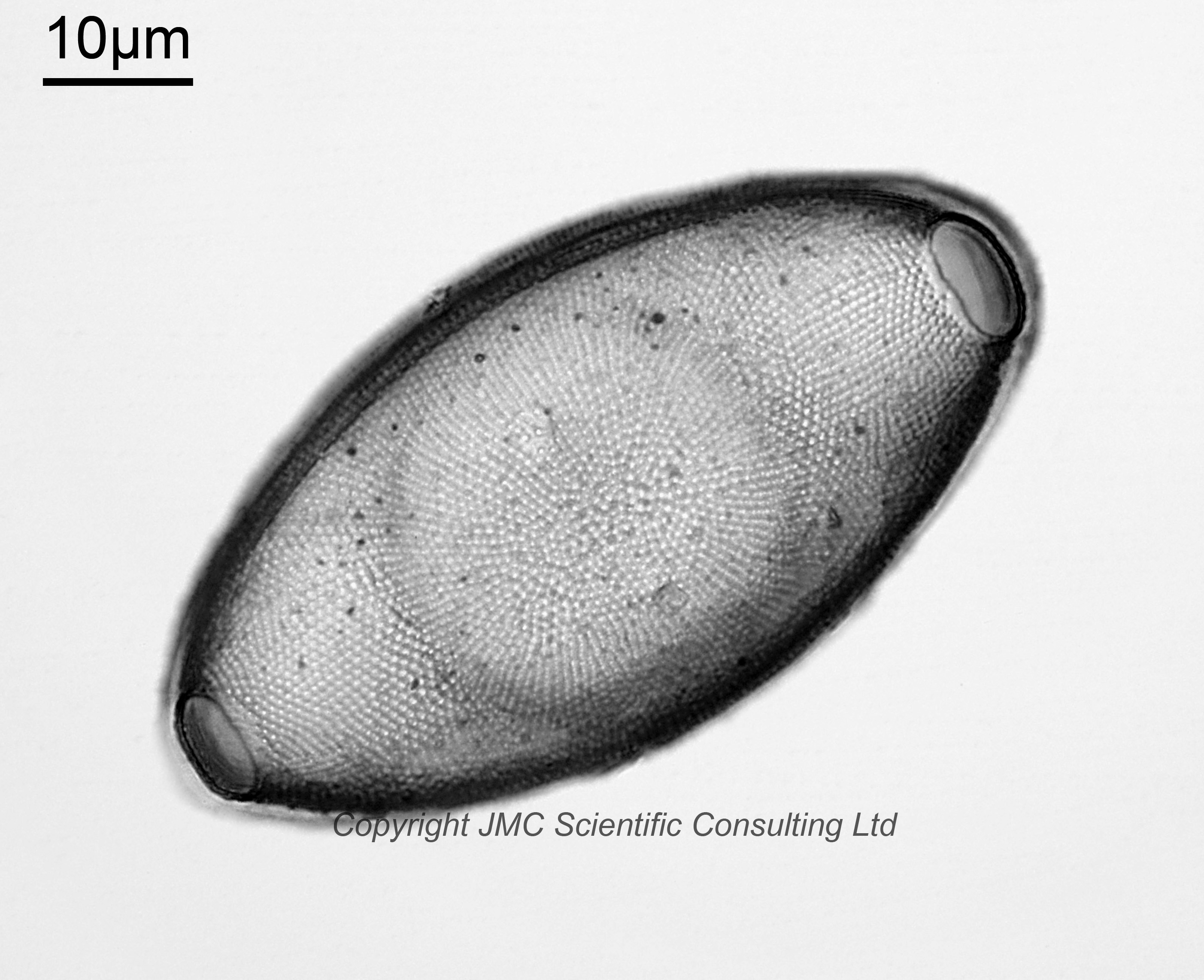
Melosira saturnalis. This is a very tentative ID, and may just be the inner portion, with the rim being missing.
Stictodiscus parallelus var. parallelus f. trigona, Stictodiscus gibbosus, Stictodiscus californicus var. areolata. I am quite confident with these ID’s. All imaged using brightfield lighting.
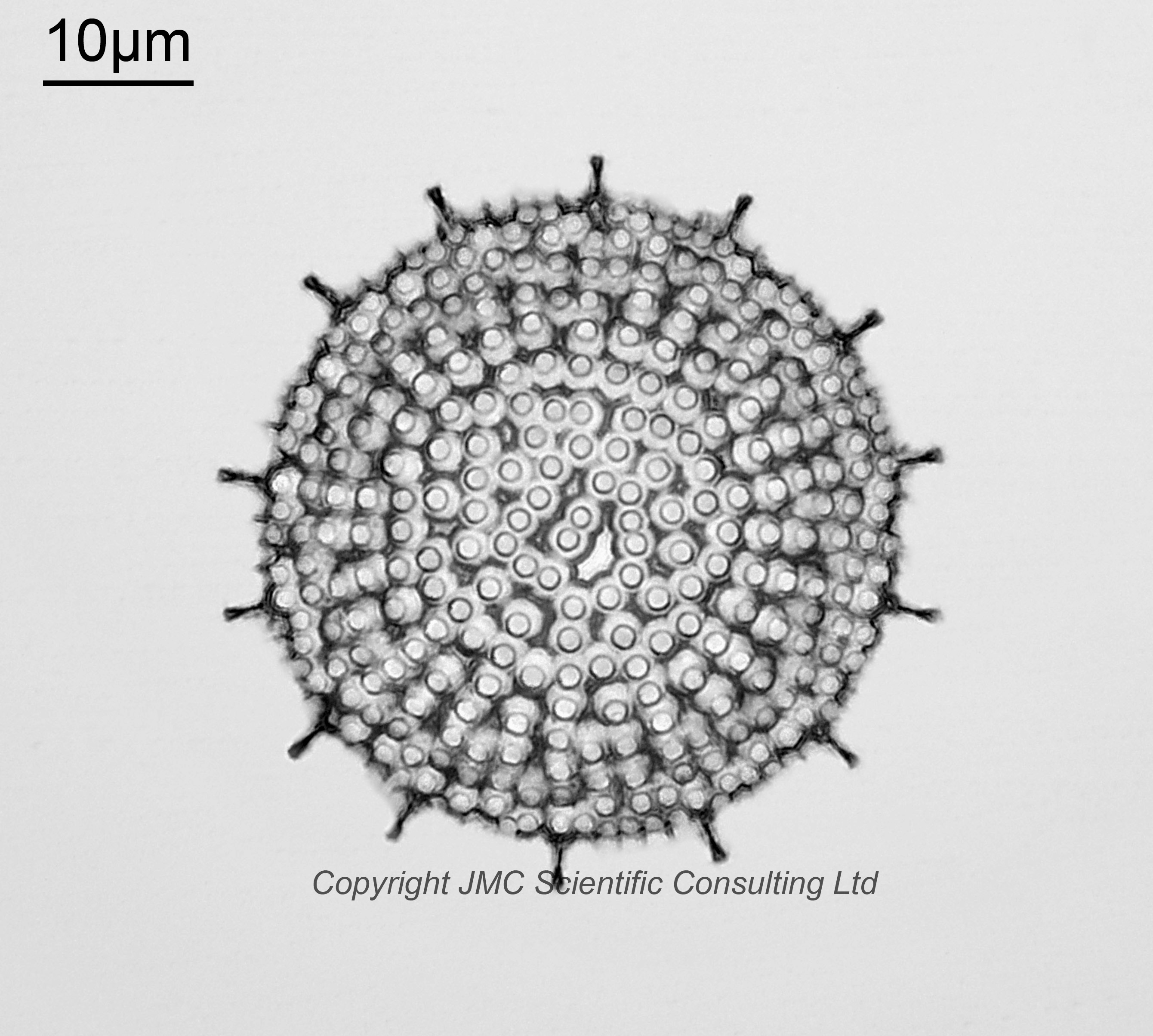
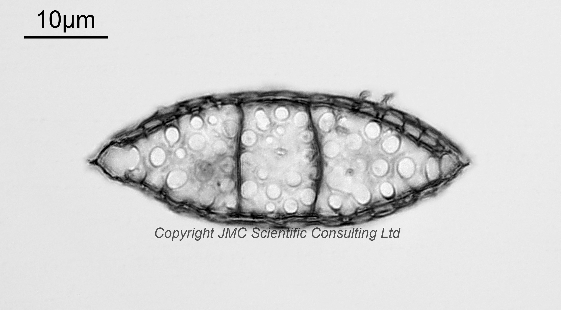
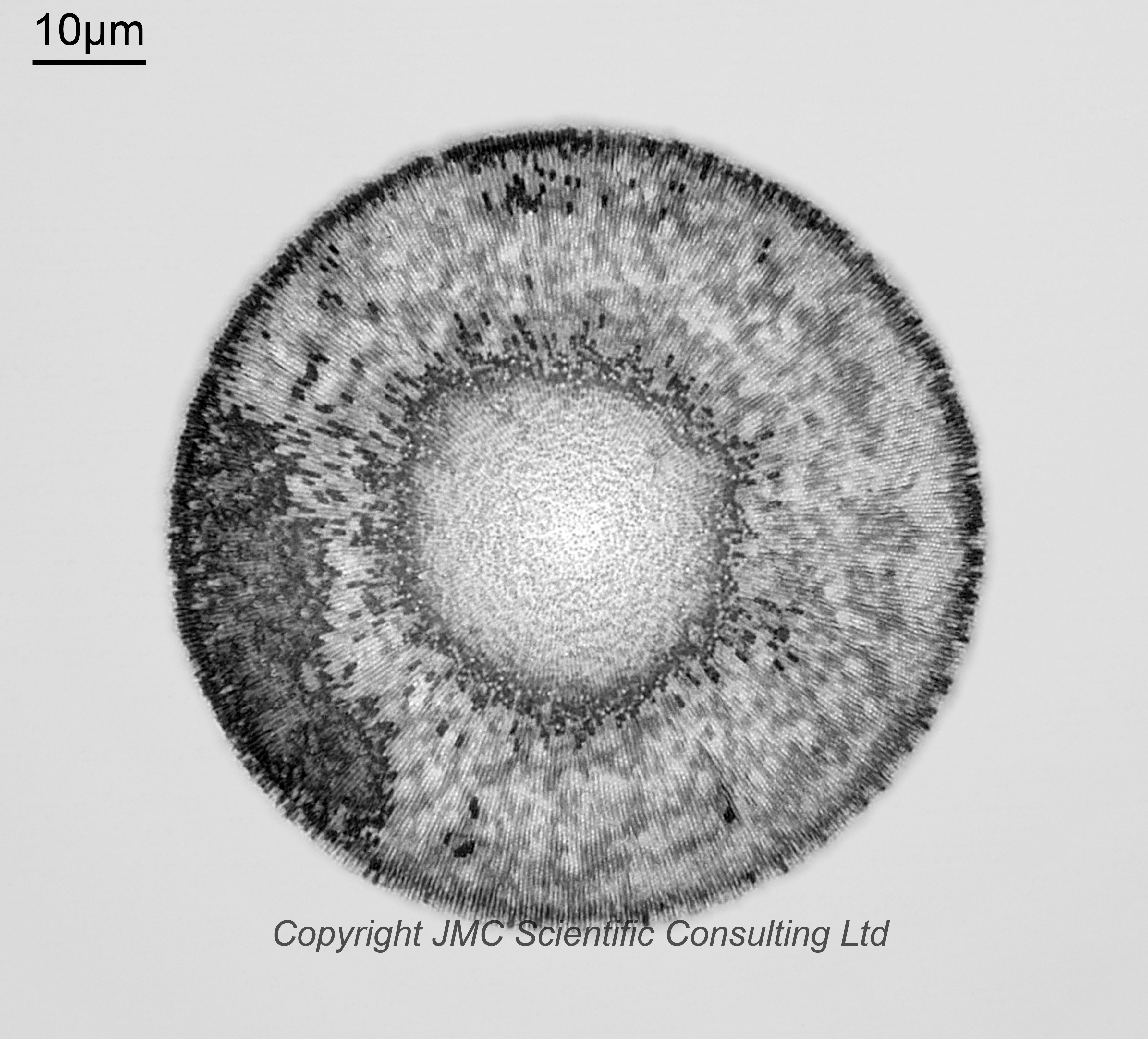
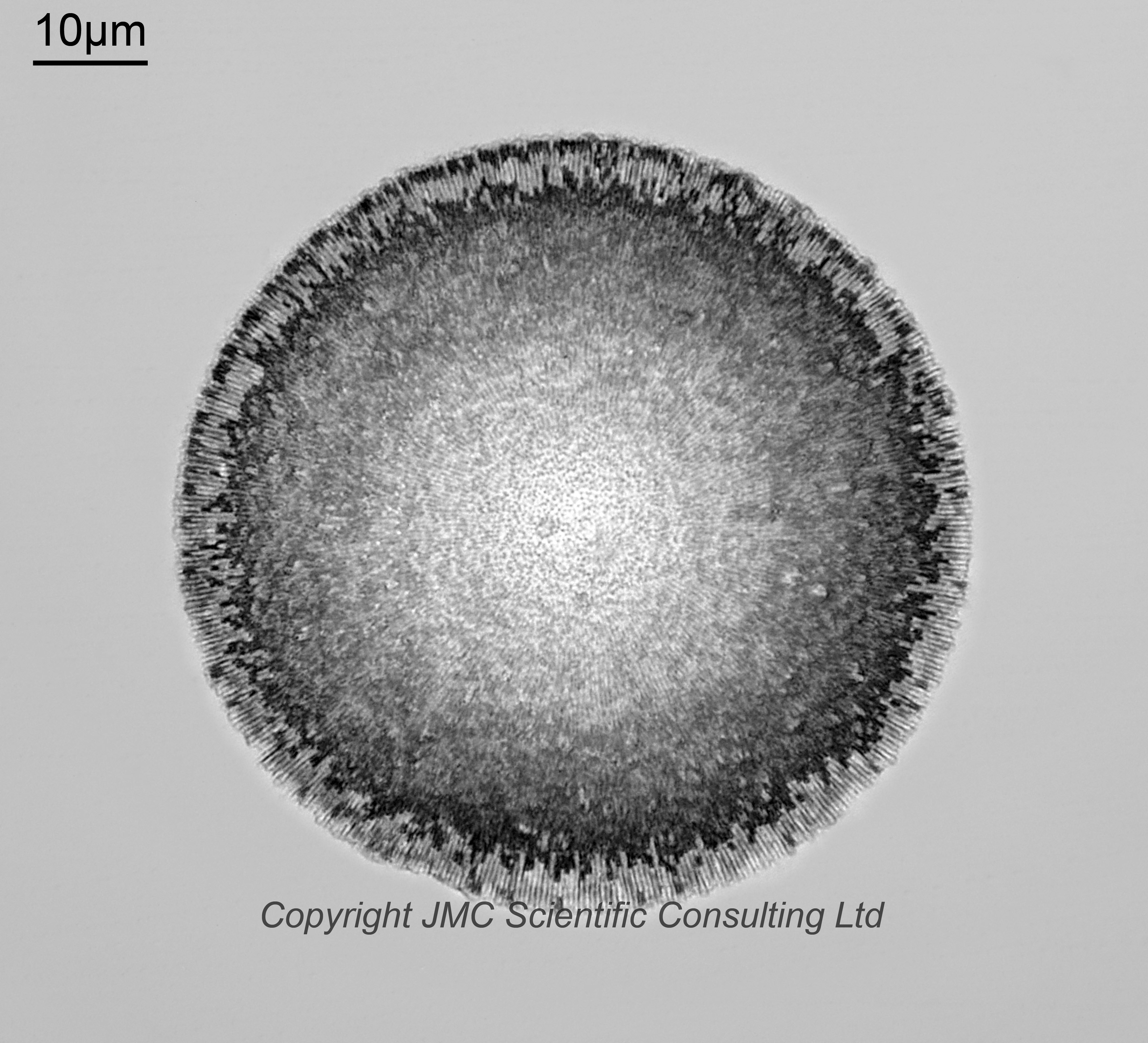
Xanthiopyxis acrolopha. I am quite confident with this name. It has also been spelt X. acrolpha and X. acrolphia. As there is a central area without the spikes I don’t think this is X. oblonga.
200-300 mesh slide
The smallest mesh size used. Again, relatively little sponge material and a higher proportion of diatoms in the mix with many either intact or almost intact. Very nice array of species, but many very small.
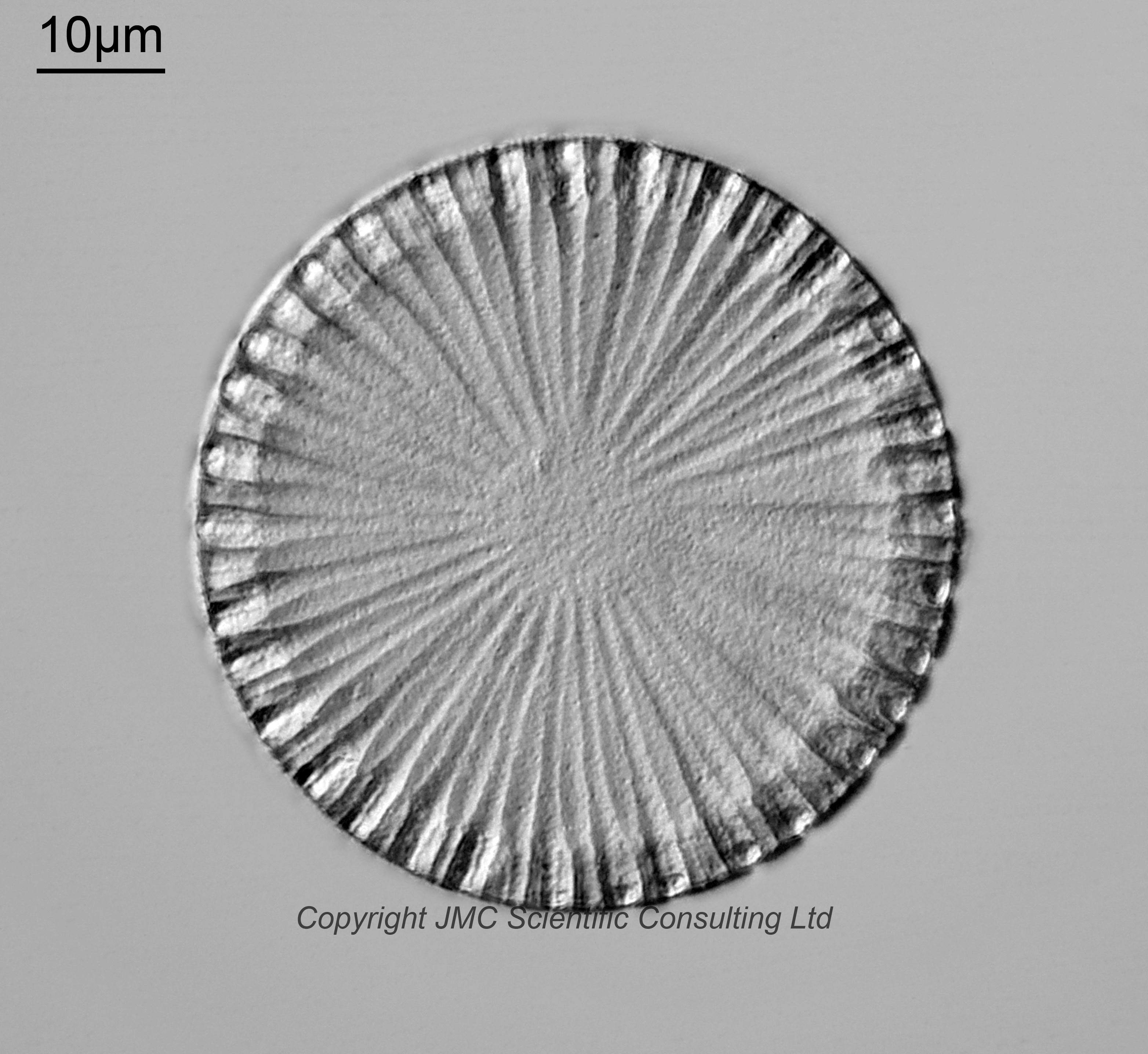
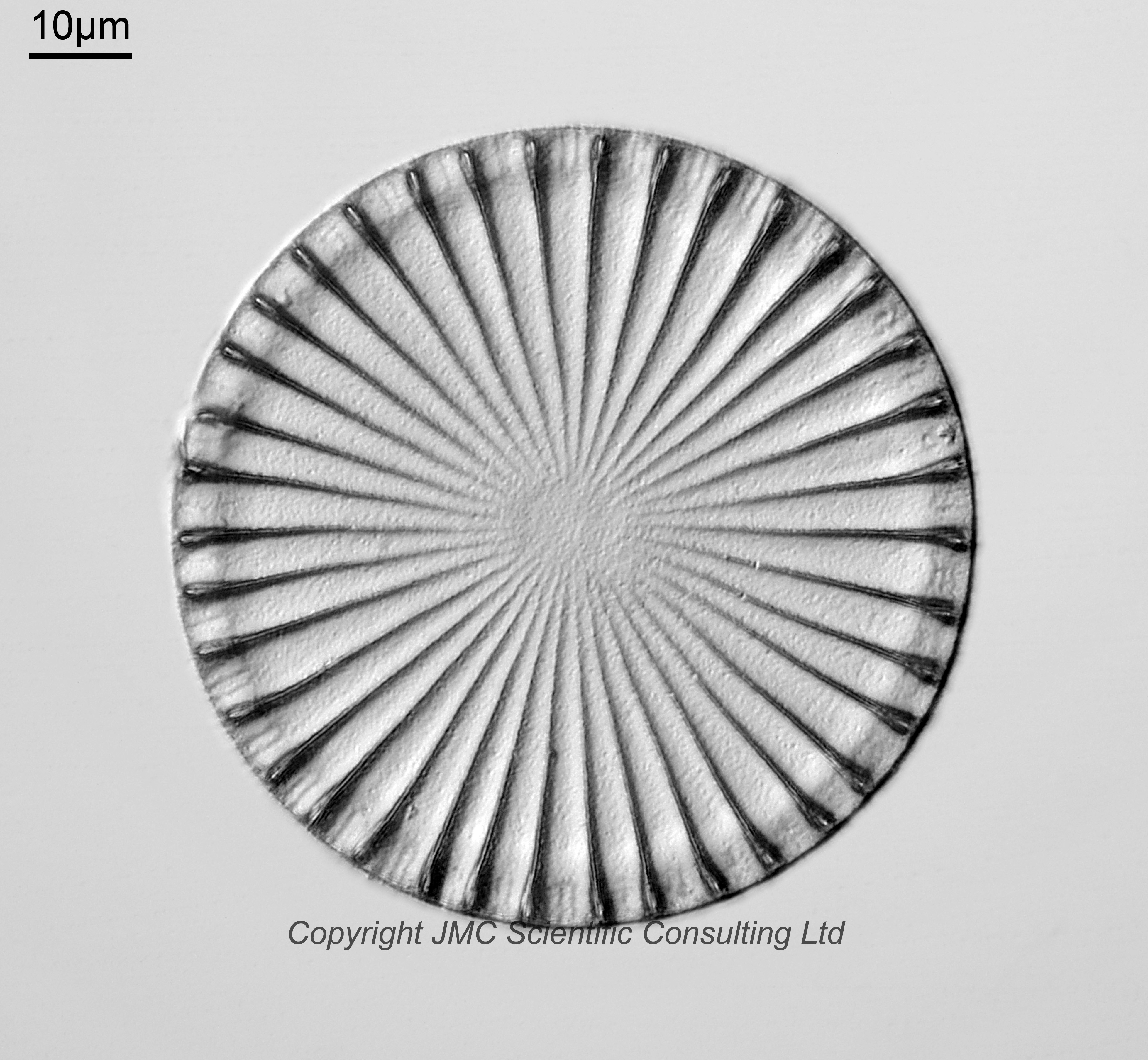
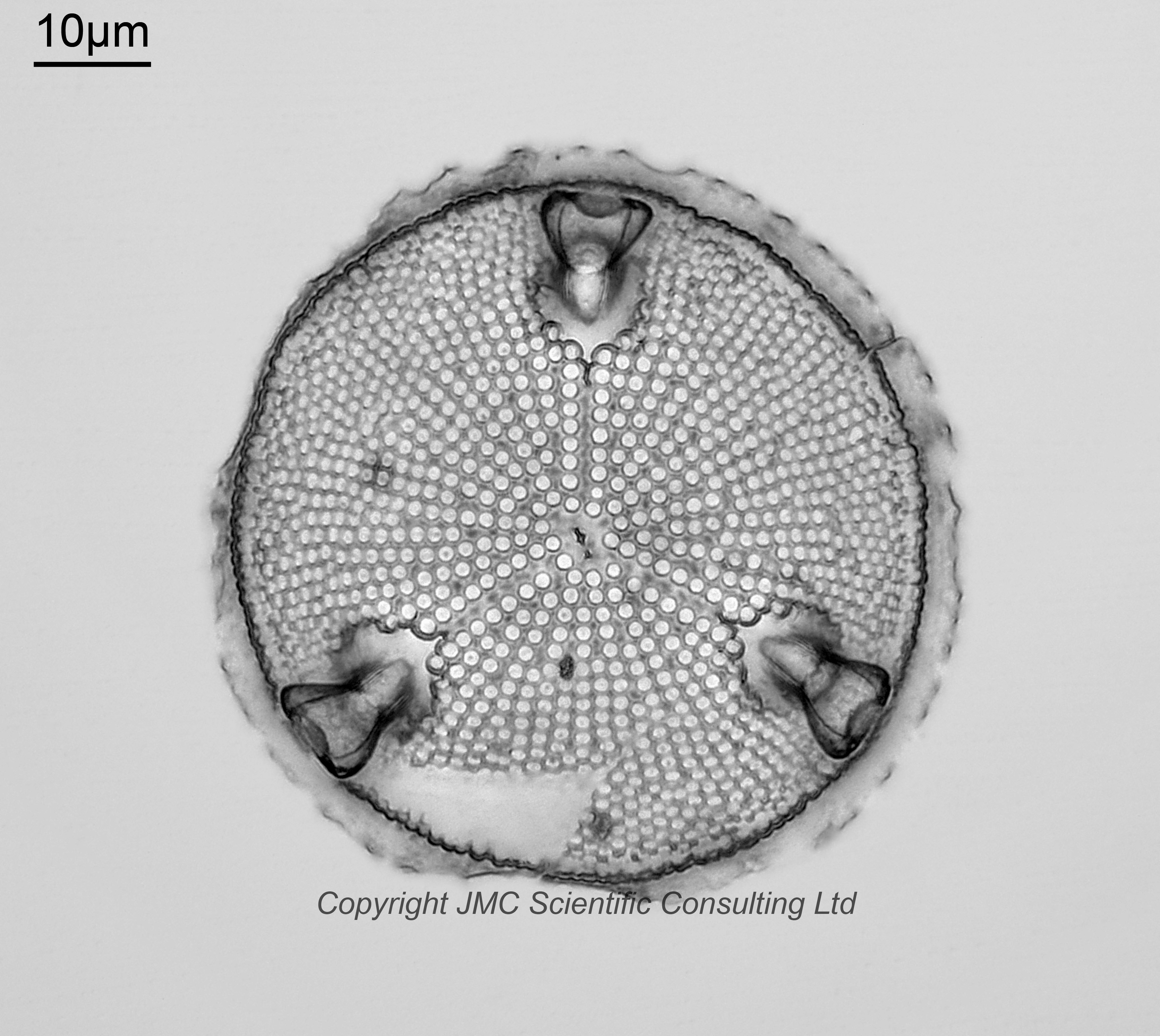
Actinoptychus splendens var. glabrata. Fairly sure on this one. Imaged using oblique lighting, which emphasizes its structure.
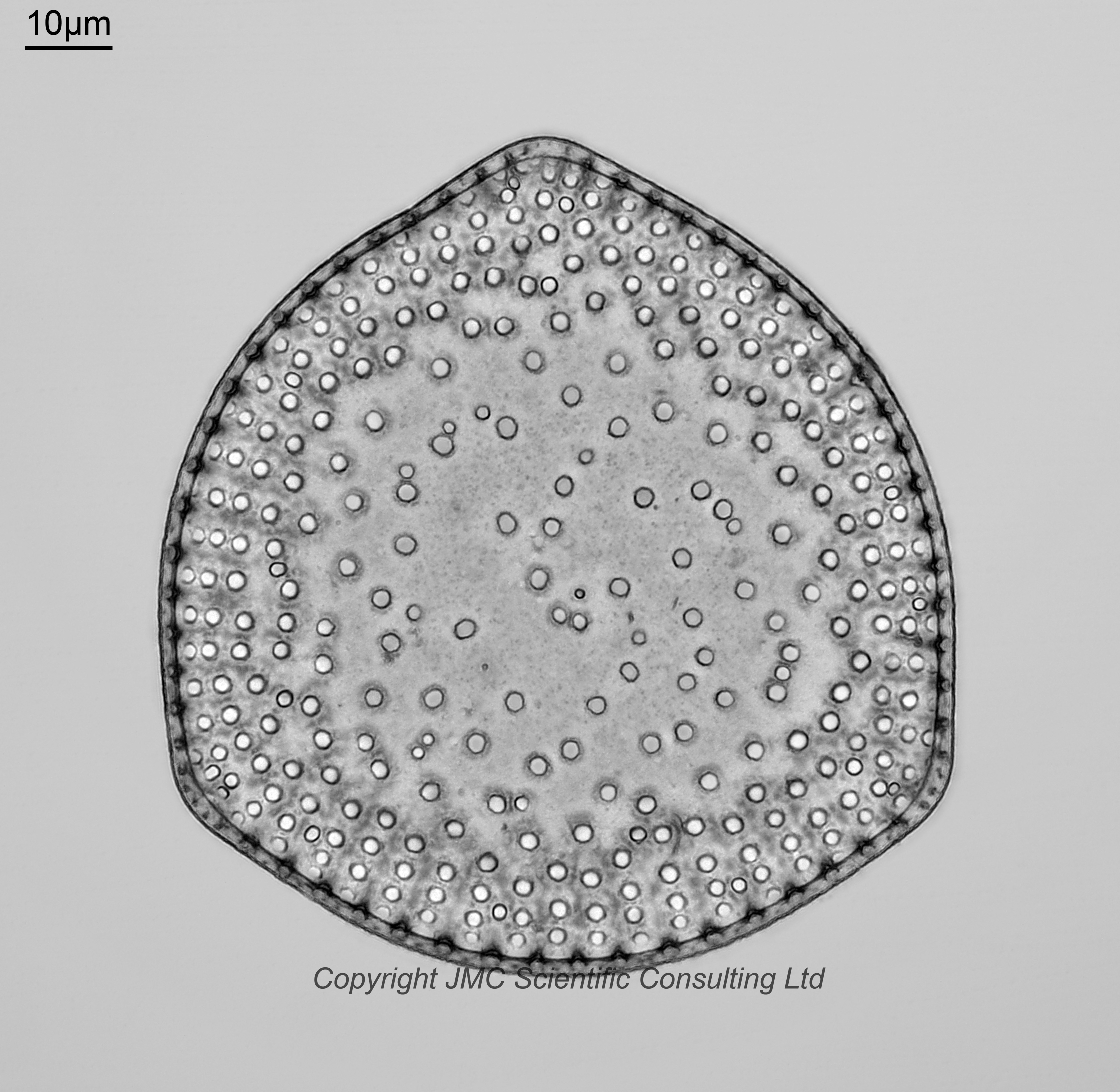
Biddulphia aurita. Again, fairly sure with the ID. Viewed from the top. Currently accepted name Odontella aurita.
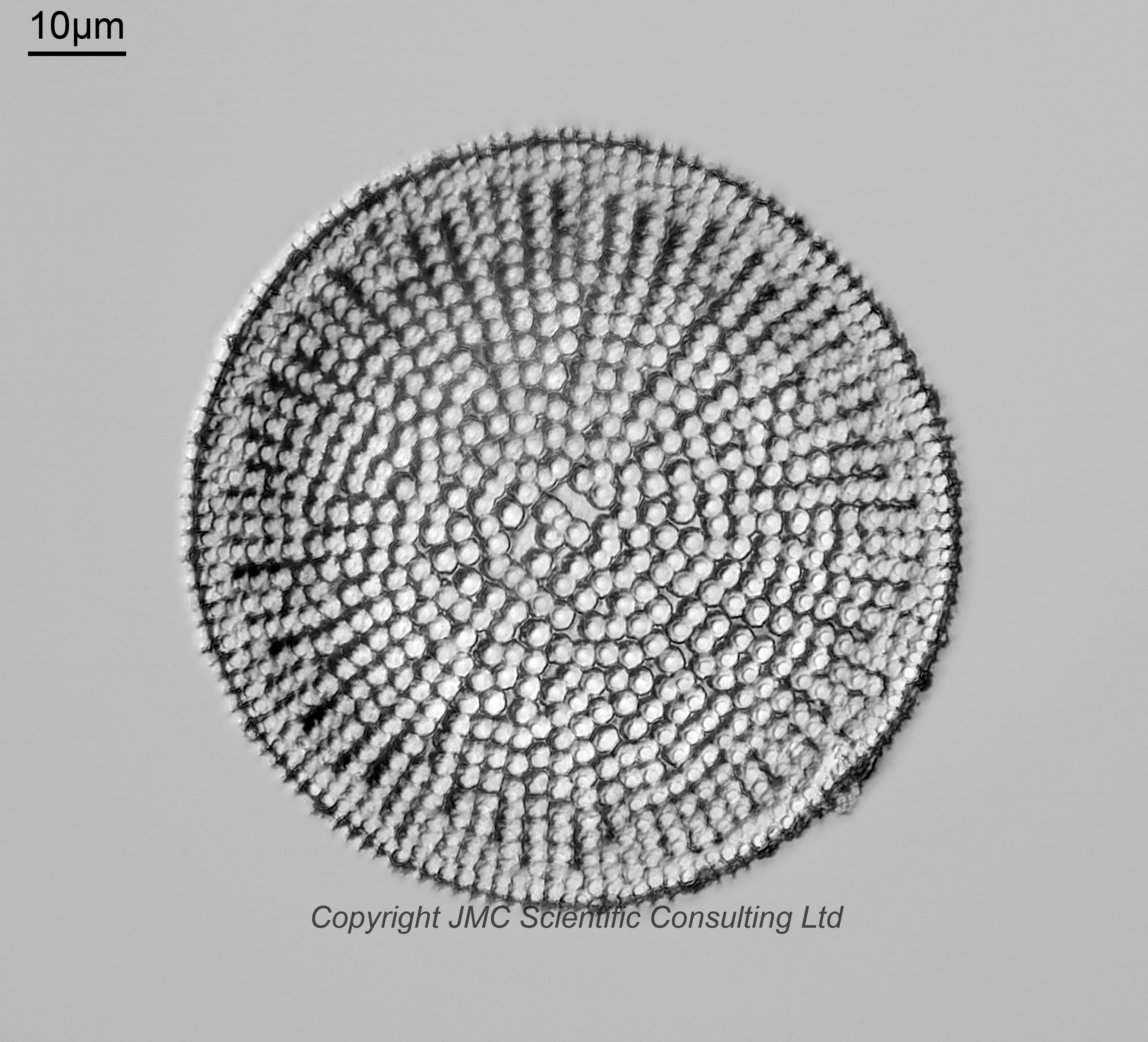
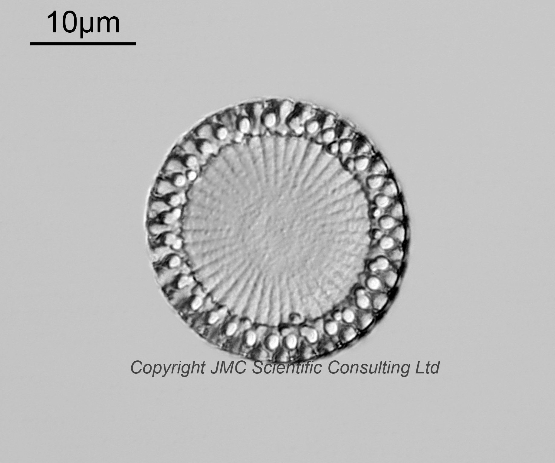
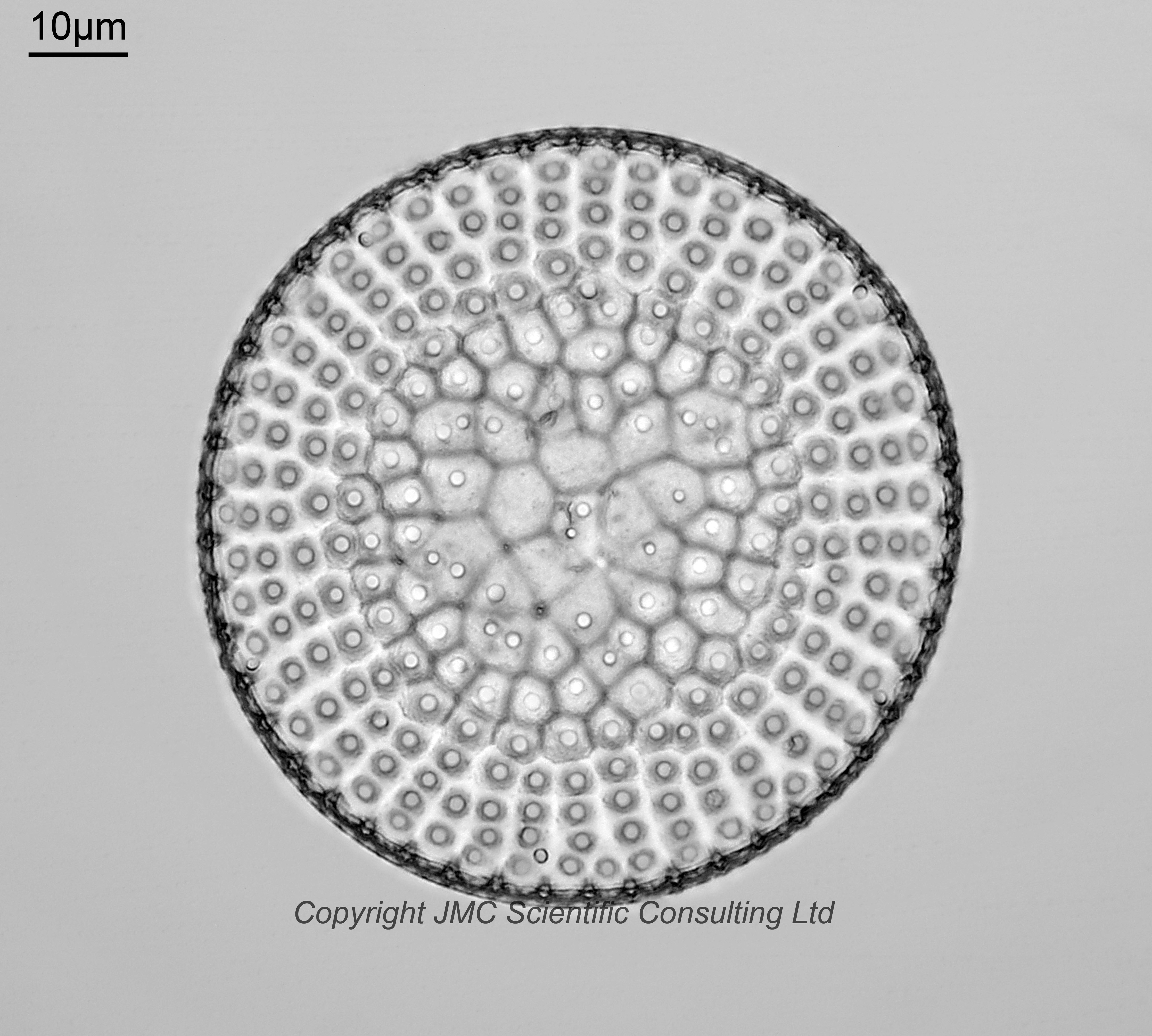
Coscinodiscus galapagensis. Less confident about this although it does look like the one in Schmidt’s Atlas, Plate 163, Figure 2. Oblique lighting.
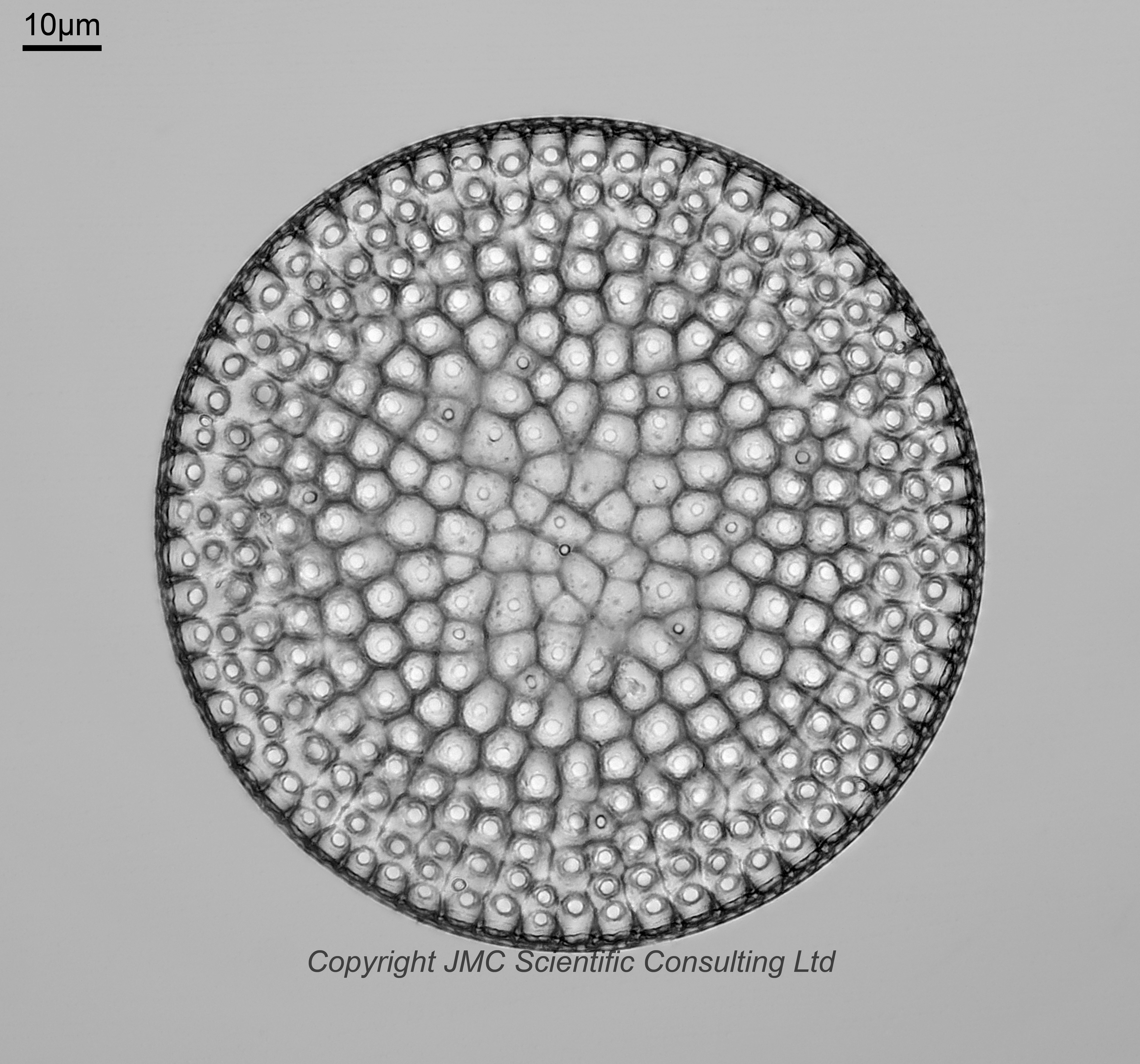
Coscinodiscus superbus. Fairly sure on this one. Oblique lighting. Couple of mentions in Schmidt’s Atlas. Plate 148, Figure 7, translation: “Oamaru (Grove), center sunken, as in Craspedodiscus; The center is lower than the edge, towards which the bowl lies bulges a bit. The highest is the wide belt surrounding the middle, in which the cells are located stand alone. Whether sp. n.? According to Witt, this form is probably related to Cestodiscus superbus from Barbadoes (Newcastle Est.) be closely related or identical.”. Also, Plate 163, Figure 8: “Oamaru, Troublesome Gully (Grove), C. superbus Hardm. MS. (Cestodiscus) var. Novae Seelandiae Grove.“.
Coscinodiscus spiniferus. Brightfield lighting.
Coscinodiscus vigilans. Single image, no stacking. Very thin diatom. Brightfield lighting. From the images in the Oamaru Diatoms website, this looks like it should be Psamodiscus nitidus, but from looking at other references for P. nitidus (for example see here), while similar in appearance I don’t think this is it, and have settled on C. vigilans.
Entogoniopsis major. Old name Triceratium majus. Brightfield lighting. Viewed from above.
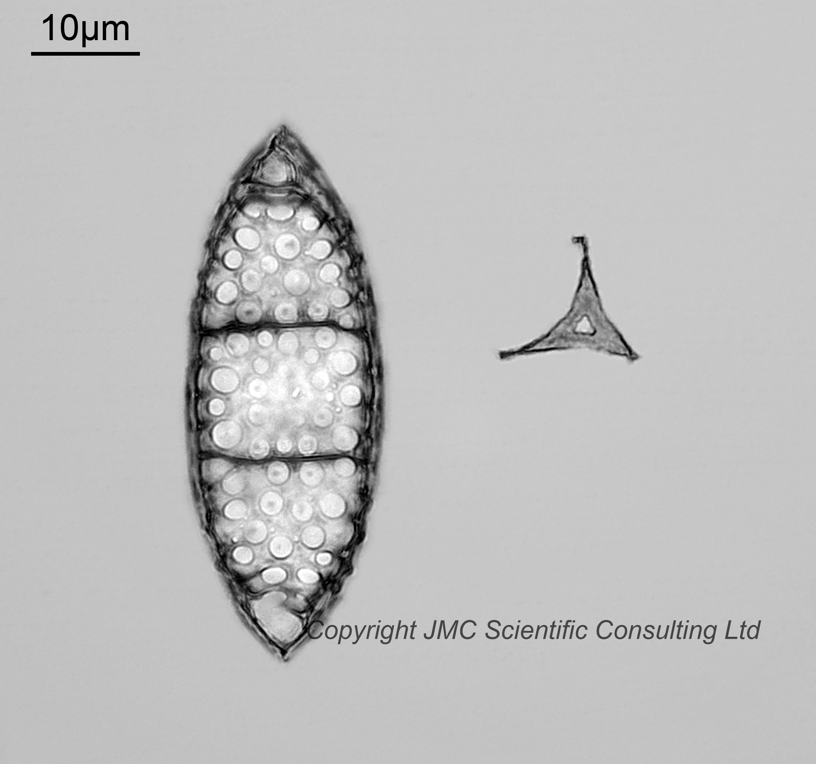
Hemiaulus polycistinorum. These two had me stumped for a while. I think they might be parts of Hemiaulus polycistinorum – the base of them with the long arms missing, and view from the top not the usual view from the side. The remains of one of the arms can just be made out on one of the images. I presume these have been damaged, probably during the filtering. Not sure as to the tiny triangular item to the right of one of the images. Imaged using brightfield lighting.
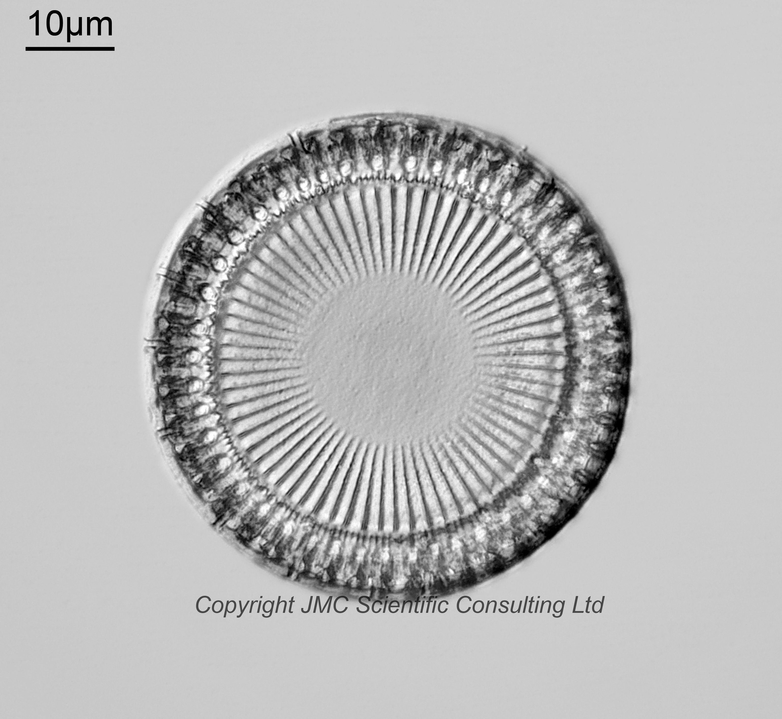
Hyalodiscus sp. I have assigned two images as Hyalodiscus of some type, perhaps H. punctatus, H. valens or H. radiatus var. striata, but I am not certain on the specific species. Both images using brightfield lighting. I put a question mark after the second image, as the central part looked to big to me. I had initially assigned this as Stellarima steinyii, as it looks very similar. But having seen the two images together, I am now thinking this may be a Hyalodiscus, however I am uncertain.
Melosira clavigera. Oblique lighting.
Melosira expectata. Very small and easily missed on the slide. Oblique lighting.
Melosira praeclara. Oblique lighting.
Paralia sulcata var. biseriata f. radiata. Oblique lighting.
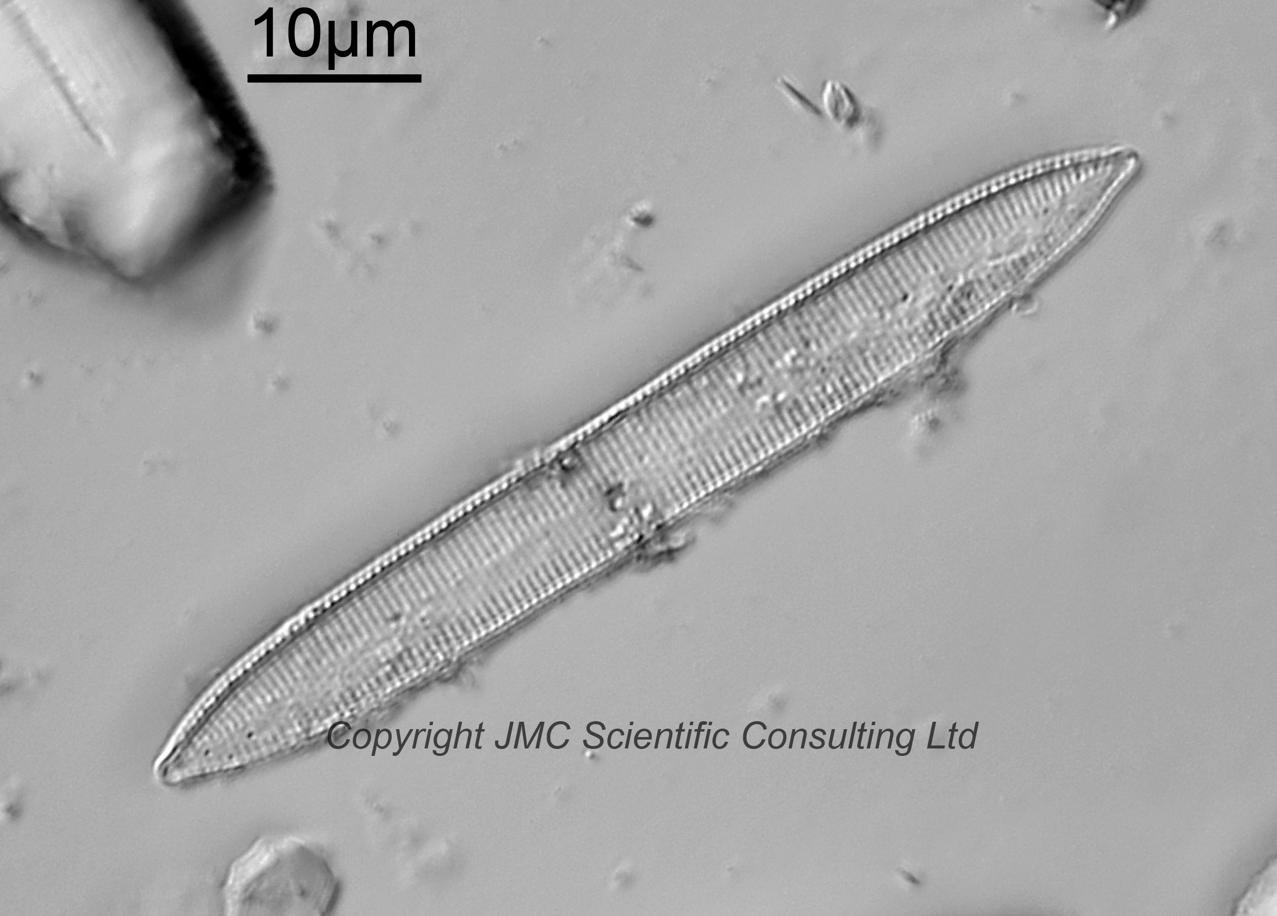
Nitzschia sp. This is a very tentative ID. Oblique lighting. Someone has mentioned it may be Tryblionella sp., although that is not one I am familiar with. Very faint and easily overlooked.
Stictodiscus californicus var. areolata. Brightfield lighting.
Stictodiscus californicus var. nitida. Viewed from underneath. Brightfield lighting.
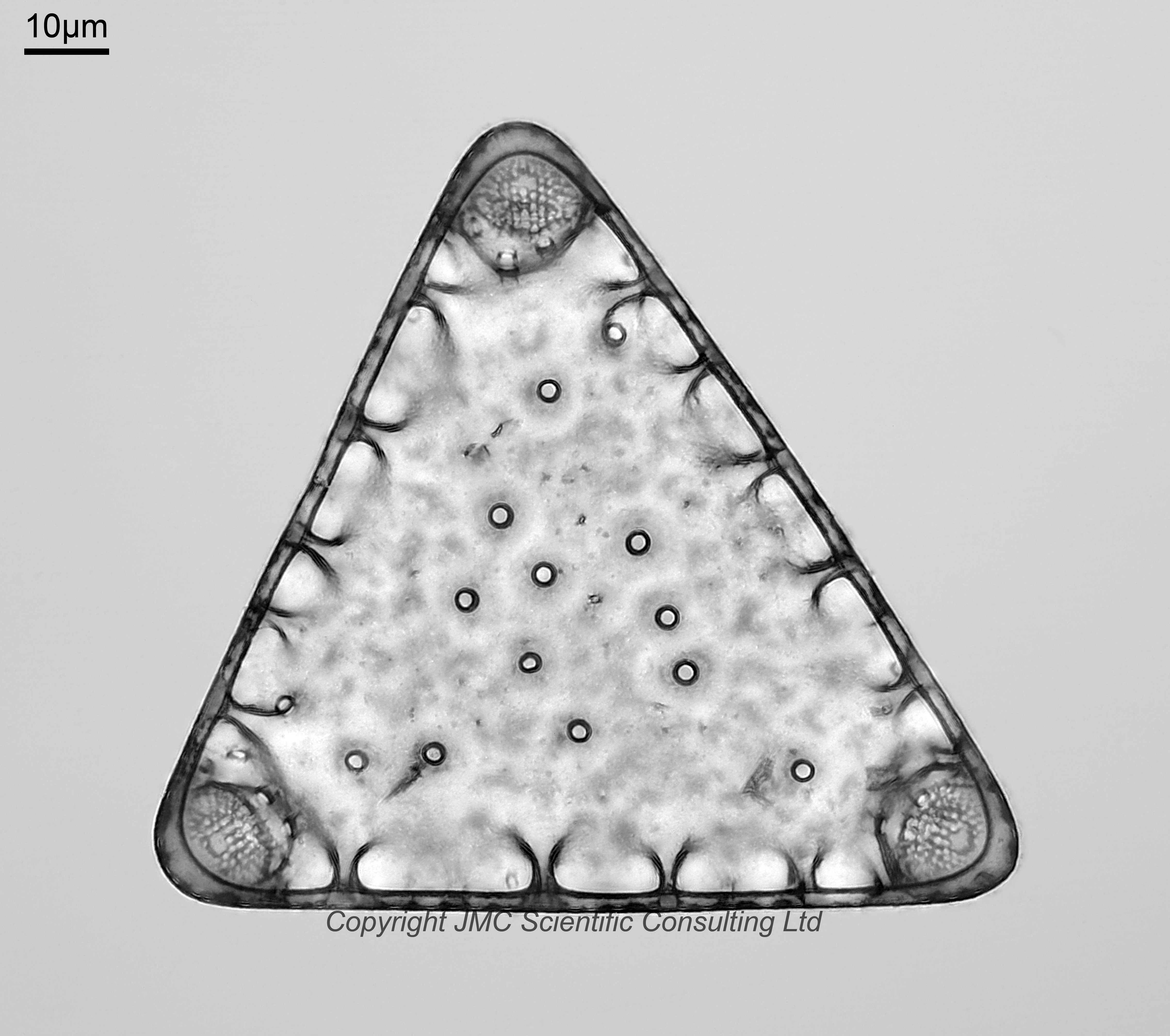
Sheshukovia castellifera. Old name Triceratium castelliferum. Broken ends. New naming discussed in Witkowski, J. (2022). Early Paleocene-Late Eocene diatoms from the Blake Nose Western North Atlantic Ocean. Nova Hedwigia, Beheift 152: [i]-v, [1]-381, incl. 2 figs, 7 tables, 156 pls. Brightfield lighting.
Triceratium anastomosans. Shown in Schmidt’s Atlas, Plate 150, Figure 10. Brightfield lighting. Looking at this I am not sure it should be a Triceratium – the apices don’t look right to me, and perhaps this should be named as a Biddulphia.
Triceratium trisulcum var. producta. Broken ends. Brightfield lighting.
Aulacodiscus sollittiianus Norman var. novazealandica. Also known as Aulacodiscus nova-zealandicus. View from above. Brightfield lighting.
Overall a fascinating set of slides and nice to see what shows up with the different filtering.