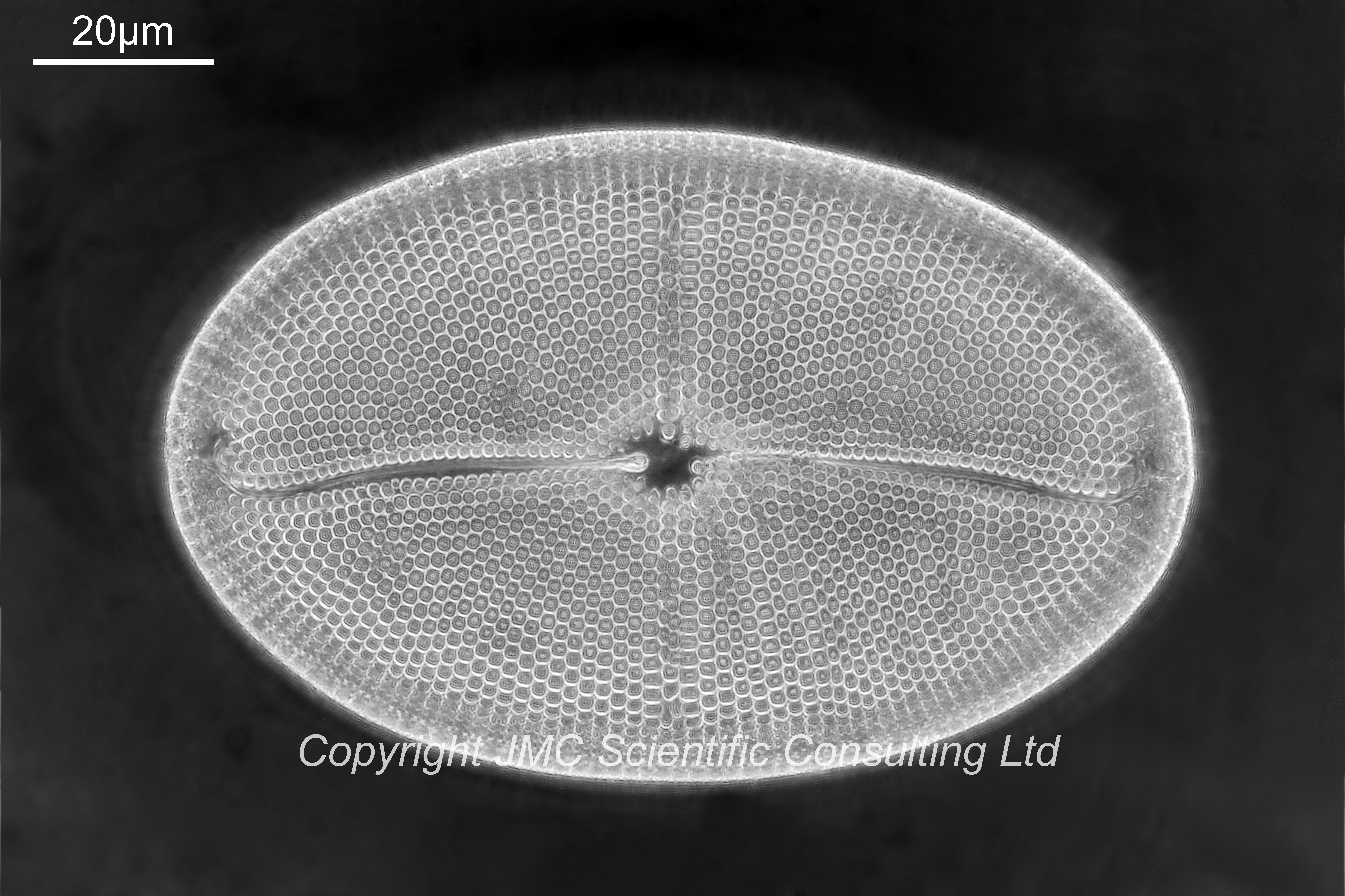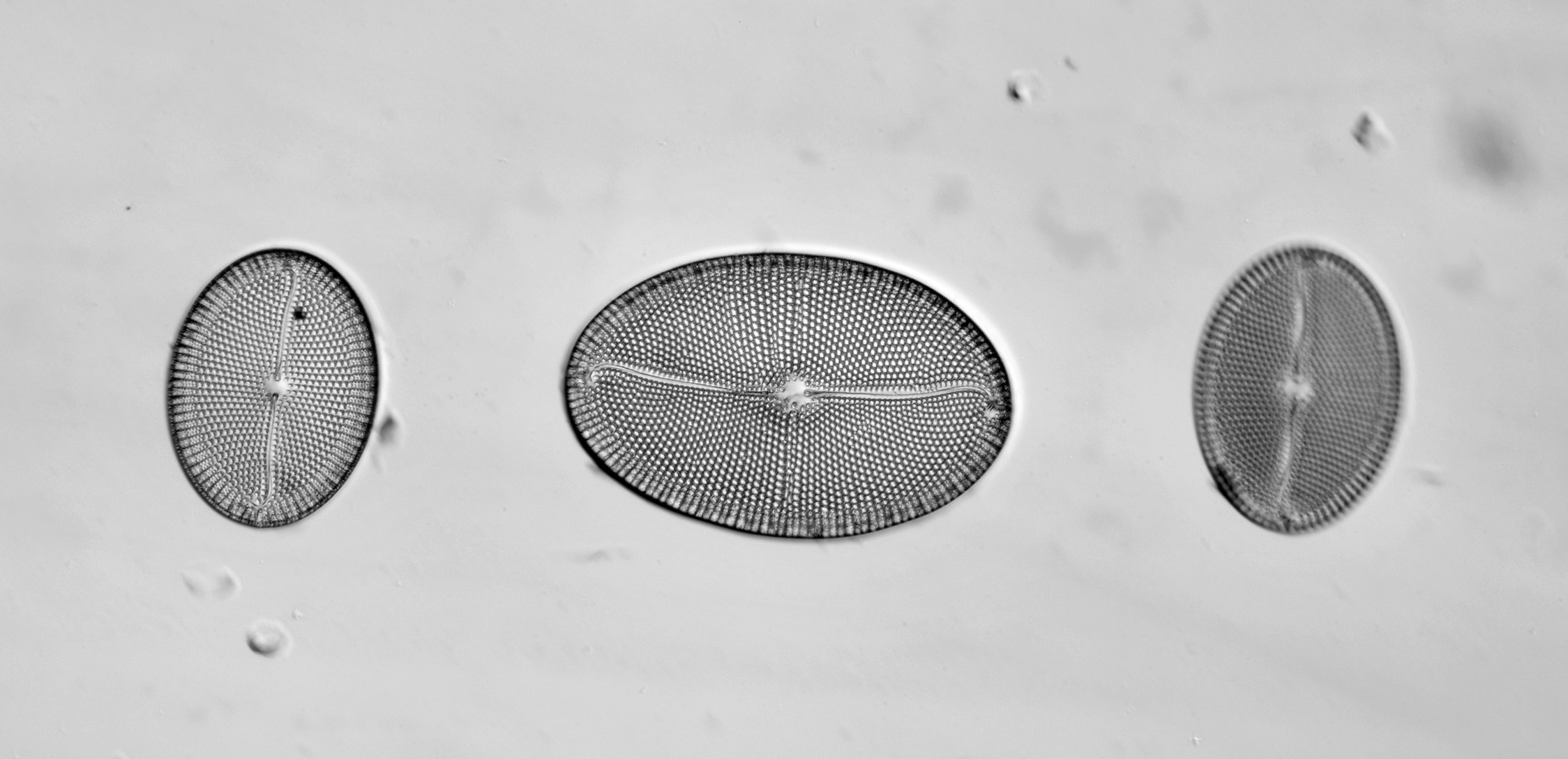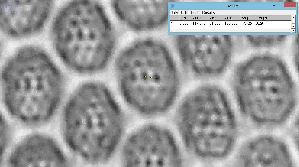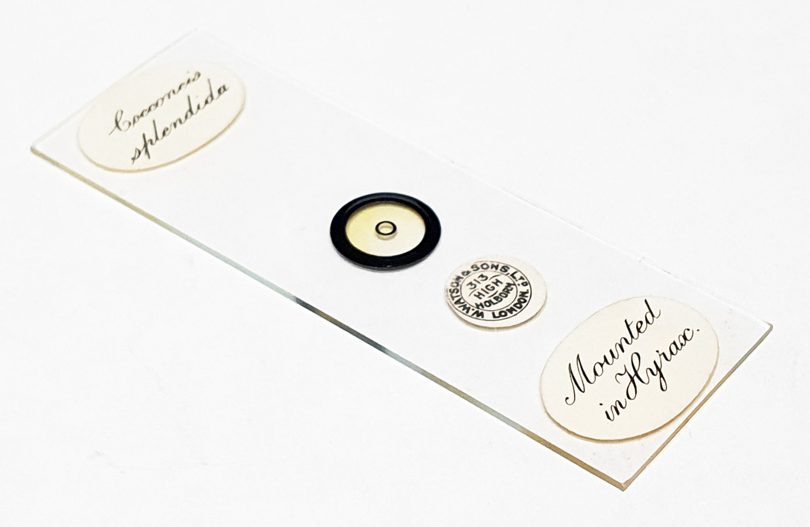


Cocconeis splendida arrangement. Three examples on the slide. Main image is of the central diatom. No collection location given. Prepared by Watson and Sons Ltd. Mounted in Hyrax. Olympus BHB microscope using 450nm LED light. Leitz 100x Pl Apo NA 0.60-1.32 objective, iris closed down slightly to produce a dark ground image. Reichert Neo 1.18/1.42 dark ground condenser, oil immersion. Nikon 2.5x CF PL photoeyepiece. Monochrome converted Nikon d850 camera. 4 images stacked in Zerene (Pmax). Small holes spaced about 291nm apart (as measured using ImageJ).