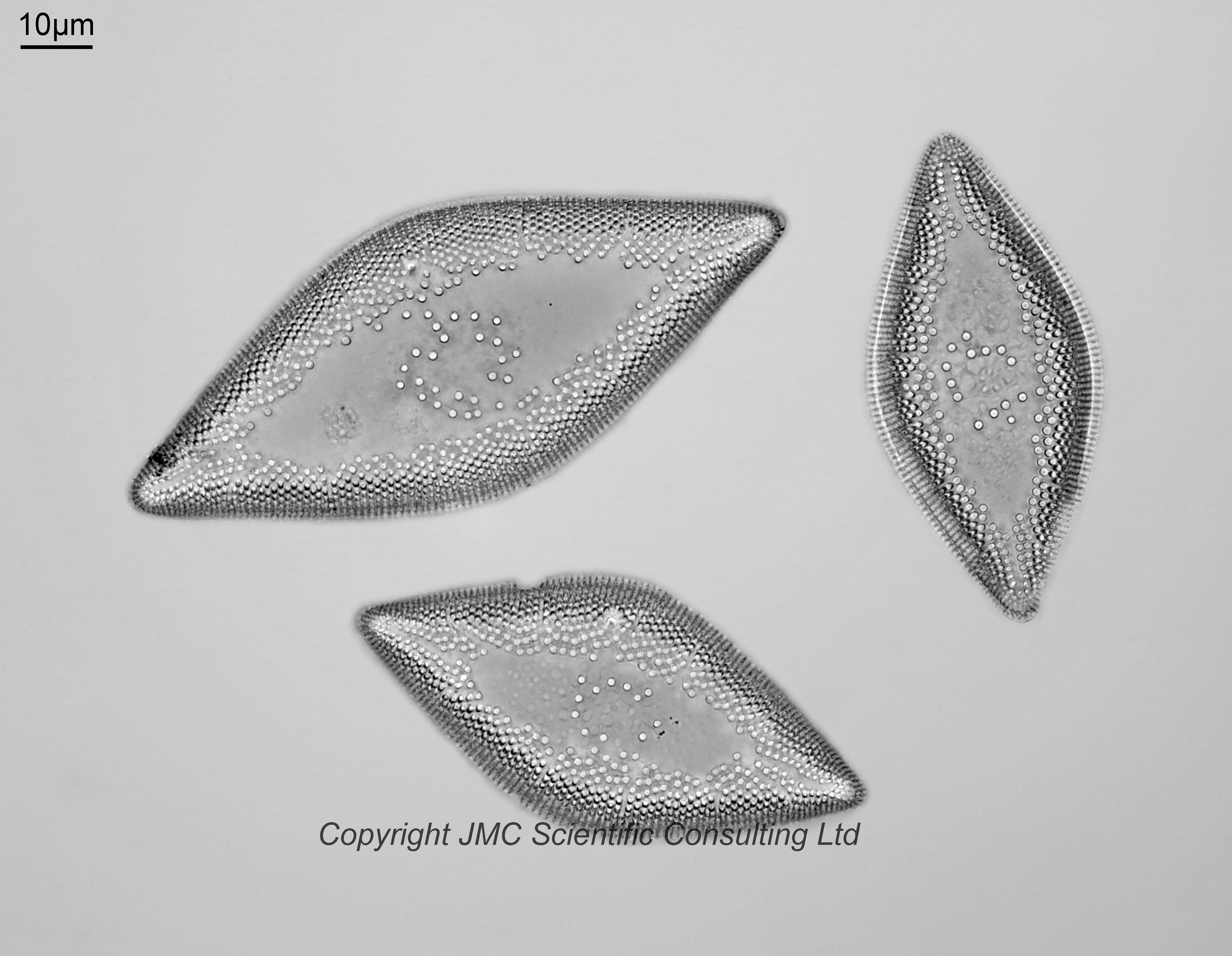
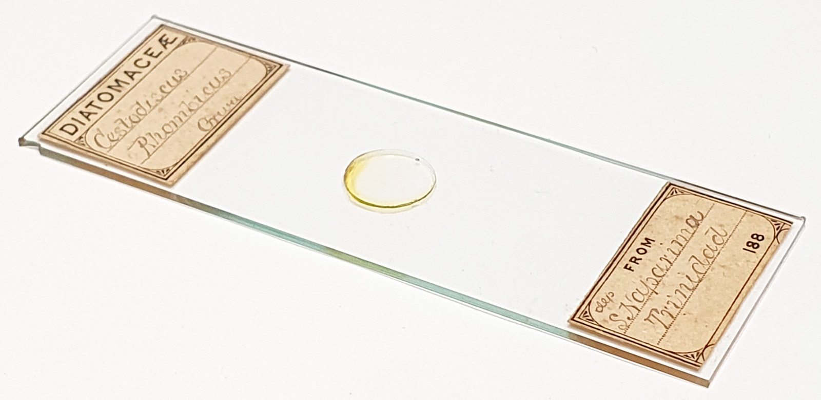
An arrangement of three Cestodiscus rhombicus from South Naparima, Trinidad. There is no makers name on the label however it is very similar to slides by WA Firth, although Barbour brothers ones are also very similar. For now I’ve put it down as WA Firth but with a question mark. Olympus BHB microscope using 450nm LED light. 63x Leitz Pl Apo NA 1.40 objective, oil immersion. Olympus Aplanat Achromat condenser, oil immersion, brightfield lighting. 2.5x Nikon CF PL photoeyepiece. Monochrome converted Nikon d850 camera. 18 images stacked in Zerene (Pmax).
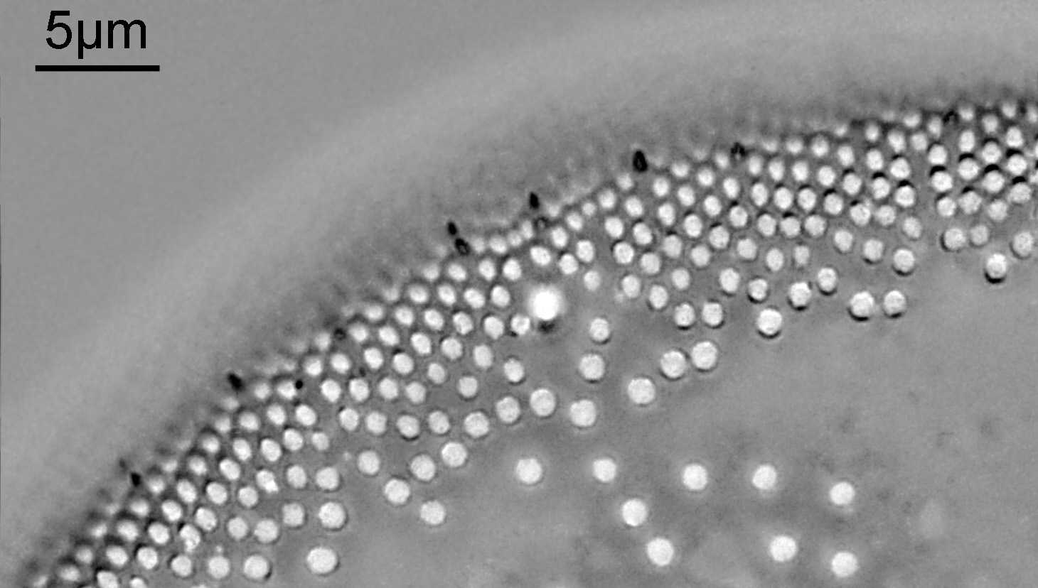
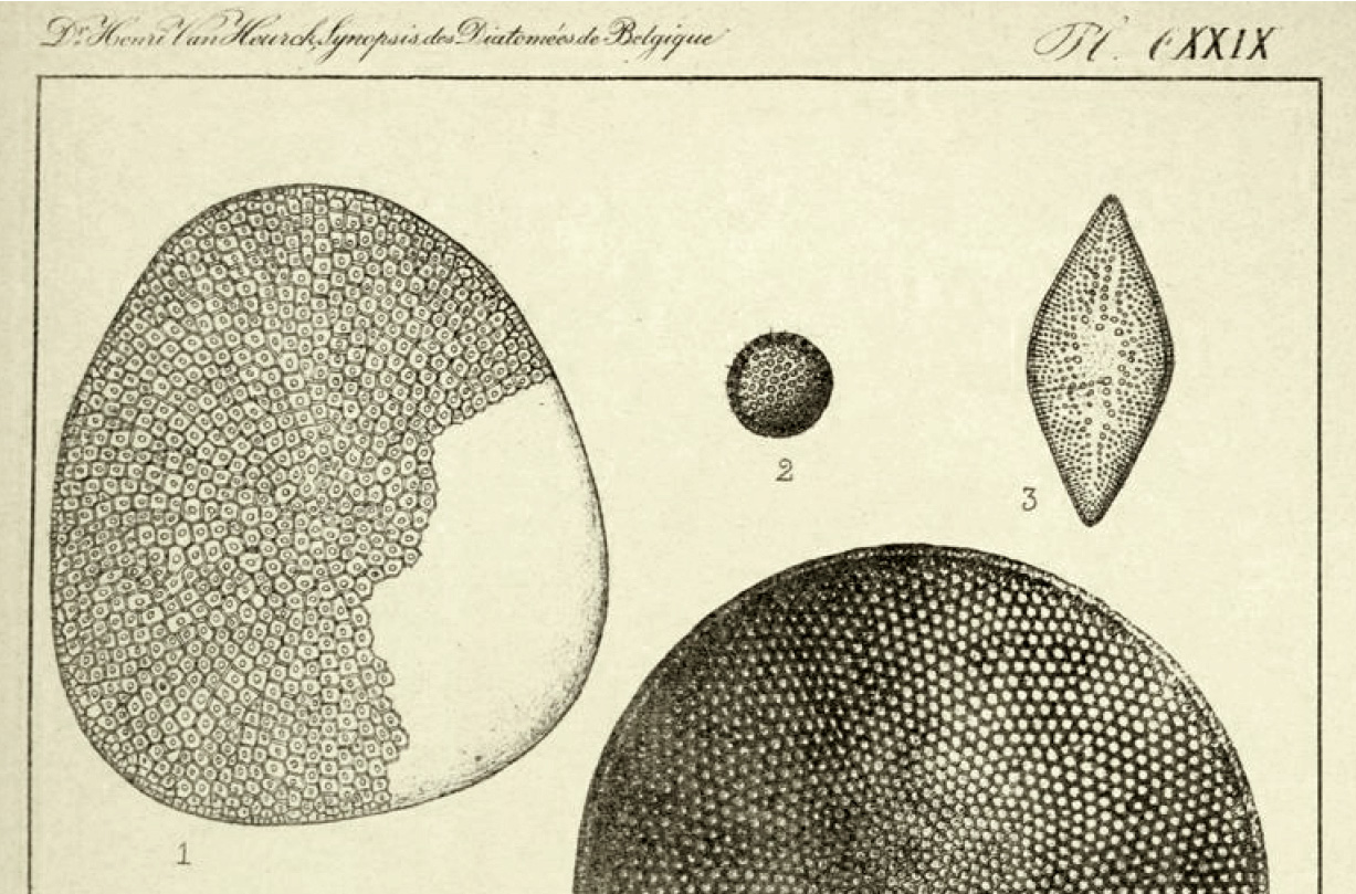
This is mentioned in Van Heurck, H. (1880). Synopsis des Diatomées de Belgique. Atlas. Ducaju & Cie., Anvers. plate 129, figure 3. A very rough Google translate of the description of it gives the following “The small spines of the sample shown are barely visible. Other specimens up to 0.10mm in length and up to 0.045mm in width have a crown of small, very visible, distant (?) spines.”. The sizes on this slide are about correct. I did see spikes in the individual frames for the scan, but they were lost in the full stack. A partial stack of 3 images makes them visible though (cropped image from the top left diatom).