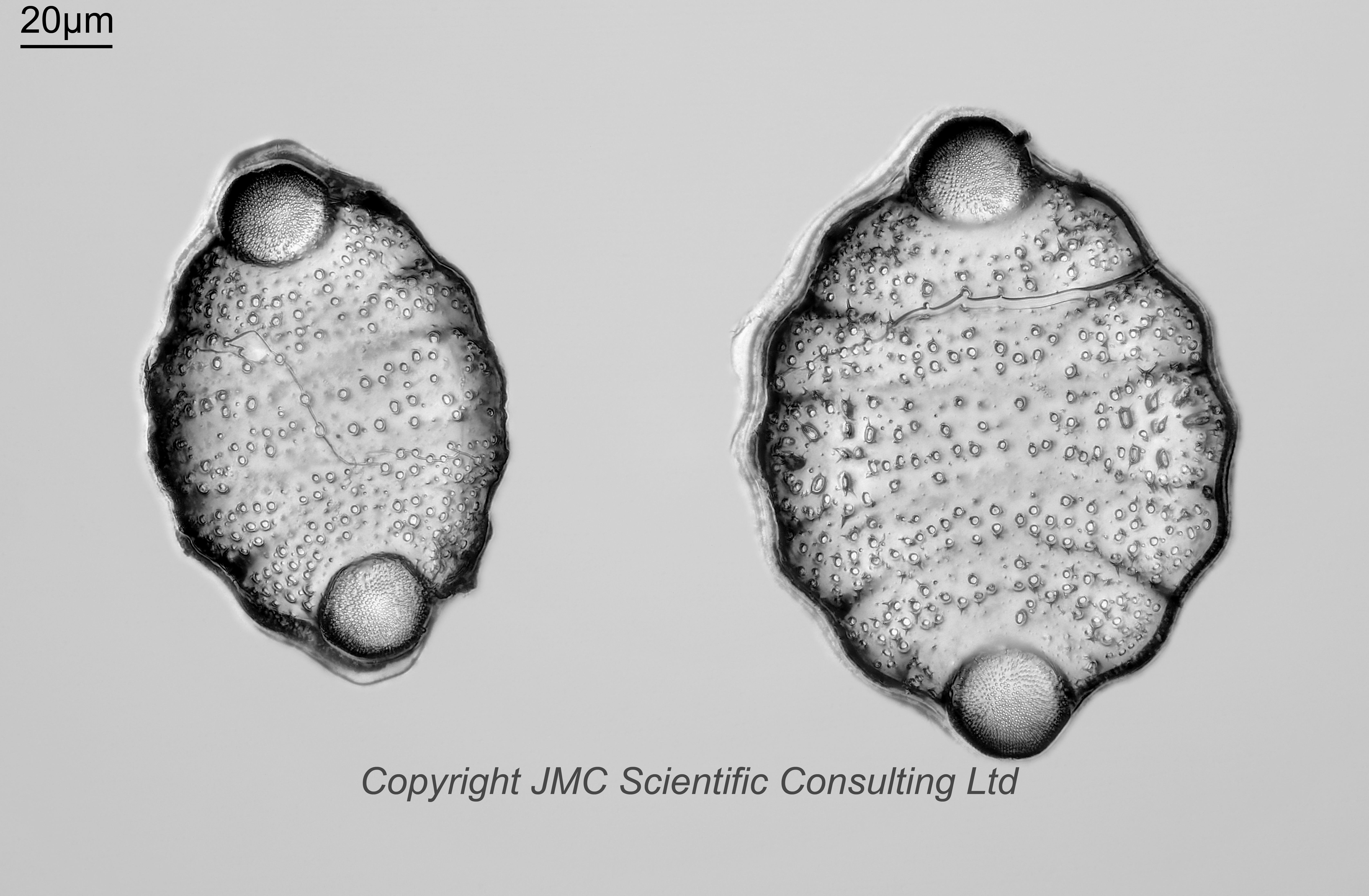
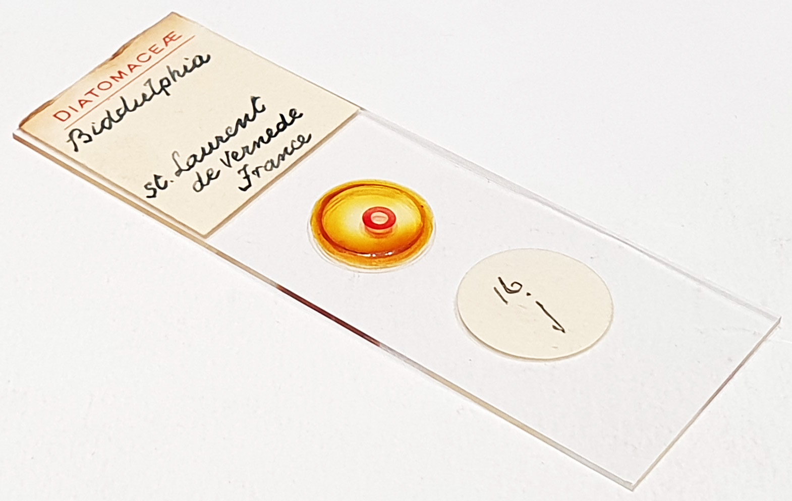
Two example of a Biddulphia sp. from St Laurent La Vernède, France (I think they are Biddulphia fortiana, more on that in a minute). No makers name, but I think this is by JA Long. Olympus BHB microscope using 450nm LED light. 40x Leitz Pl Apo NA 1.00 objective, oil immersion. Olympus Aplanat Achromat condenser, oil immersion, oblique lighting. 2.5x Nikon CF PL photoeyepiece. Monochrome converted Nikon d850 camera. 65 images stacked in Zerene (Pmax).
Biddulphia fortiana Tempère. Original description: Forti, A. (1913). Contribuzioni Diatomologiche XIII. Diagnoses Diatomacearum quarumdam fossilium italicarum. Atti del Reale Istituto Veneto di Scienze Lettero ed Arti, 72(2): 1535-1701. Page 1601, Plate 8, Figures 1,5,20,24.
My ID for this one comes mainly from Sims, P.A., Ashworth, M.P., Theriot, E.C. & Manning, S.R. (2022 ‘2023’). Molecular and morphological analysis of Biddulphia sensu lato: a new diagnosis of Biddulphia, with a description of the new genera Biddulphiella and Neobrightwellia. Marine Micropaleontology 178(102186): 1-21, 21 figs. Page 9, Figure 4.