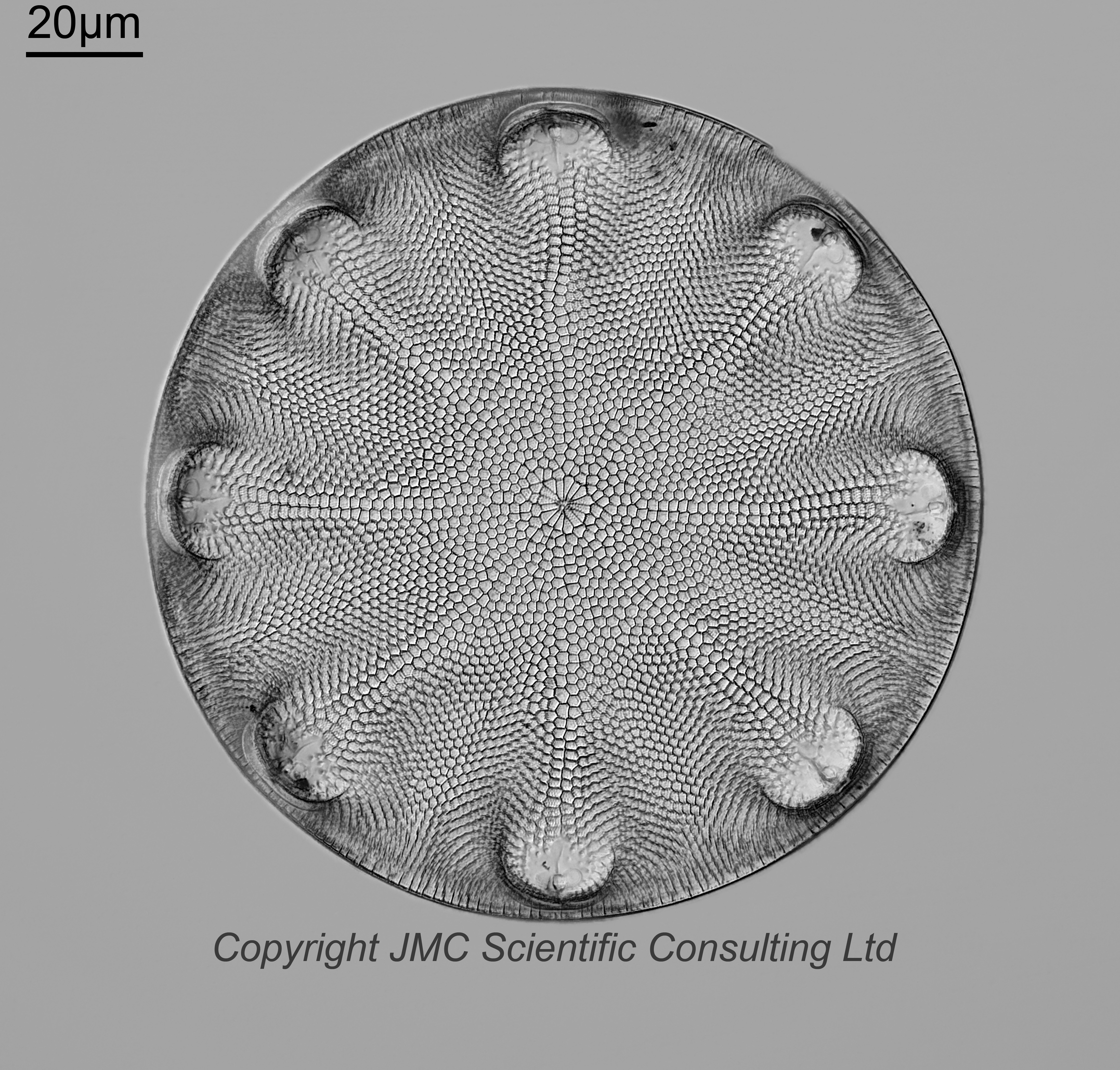
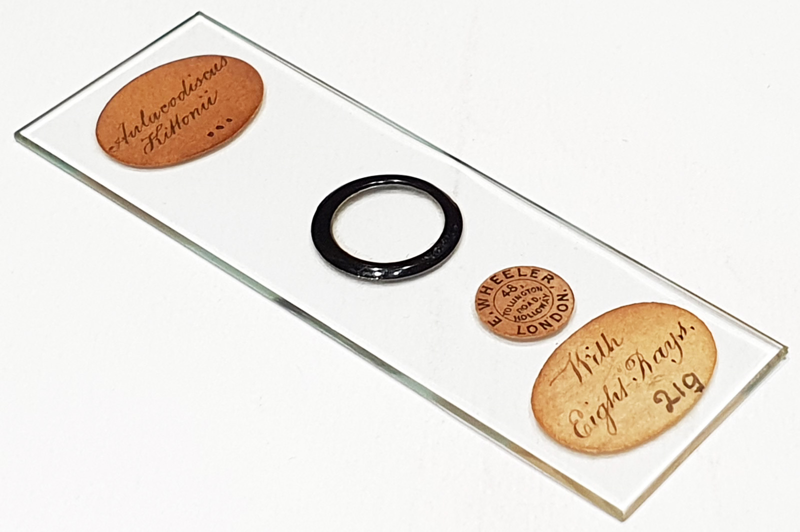
Aulacodiscus kittonii ‘with 8 rays’. Single example on the slide. 8 processes. A ‘three dot’ slide. Prepared by Edmund Wheeler. Olympus BHB microscope using 450nm LED light. 40x Leitz Pl Apo 1.00 objective, oil immersion. Olympus Aplanat Achromat condenser, oil immersion, oblique lighting. 2.5x Nikon CF PL photoeyepiece. Monochrome converted Nikon d850 camera. 85 images stacked in Zerene (Pmax). Couldn’t get close enough to use the 63x Leitz objective, as the coverslip ring is quite tall.
Aulacodiscus kittonii Arnott ex Ralfs 1861. Published in: Pritchard, A. (1861). A history of infusoria, including the Desmidiaceae and Diatomaceae, British and foreign. Fourth edition enlarged and revised by J. T. Arlidge, M.B., B.A. Lond.; W. Archer, Esq.; J. Ralfs, M.R.C.S.L.; W. C. Williamson, Esq., F.R.S., and the author. pp. [1]-xii, [1]-968, 40 pls [I-XV]. London: Whittaker and Co.. Page 844, Plate 8, Figure 24.
Seems to be a very rare example – 8 (actually 6, 7 or 8) processes were not observed in the paper by Robert W. Holmes & Albert Mahood (1980), Aulacodiscus kittonii Arnott – distribution and morphology on the West Coast of the United States, British Phycological Journal, 15:4, 377-389, DOI: 10.1080/00071618000650381).
Also see Rivera, P.; Cruces, F.; Gallardo, V. A. First Report of a Surf Zone Diatom Accumulation in the Eastern South Pacific: Aulacodiscus Kittonii Arnott Ex Ralfs (Bacillariophyta) As the Dominant and Exclusive Species. Gayana Bot. 2016, 73, 1-8. In the discussion they stated; “The number of labiate processes on each cell is a variable feature. With light microscopy, we observed normally valves with four, five, and six processes, and rarely, cells with eight or ten processes were also found (Fig. 3. D-E). As valves with those characteristics had not been found in our previous SEM observations of the same samples, clean material dried on cover glasses at room temperature was observed under a light microscope and valves with eight or ten processes were encircled with an special diamond objective. Their observation with scanning microscopy showed valves only with four (Fig. 4. E), or five processes (Fig. 4. F), proving that the “eight” and “ten” processes valves observed at low magnification with light microscopy were an optical effect produced by two un-separated valves, each one with four or five processes respectively. The same opinion was expressed by Holmes & Mahood (1980).”
With the one on this slide, this is not two valves, this is one valve with 8 processes. Very unusual.
For an example of one with 7 processes see here.