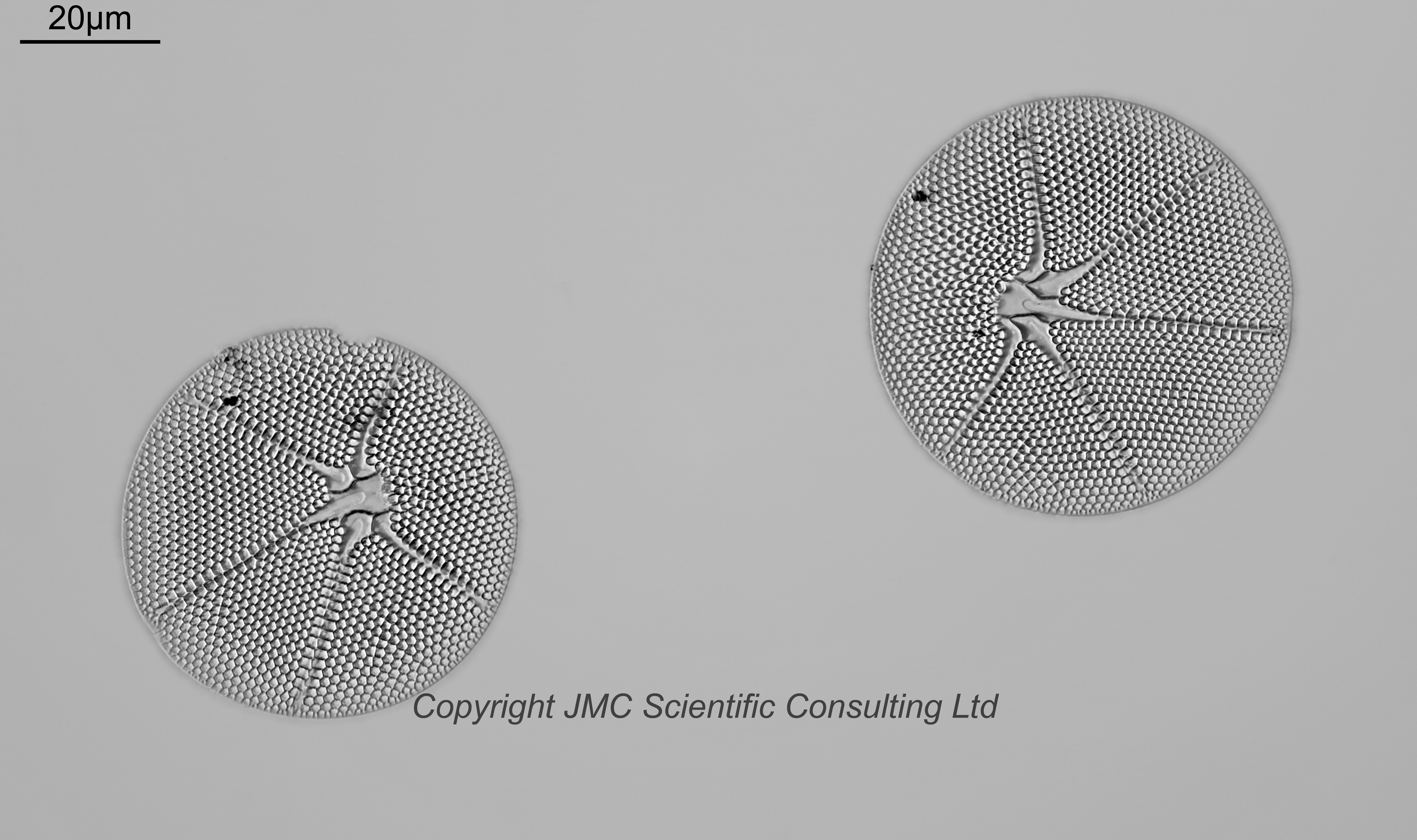
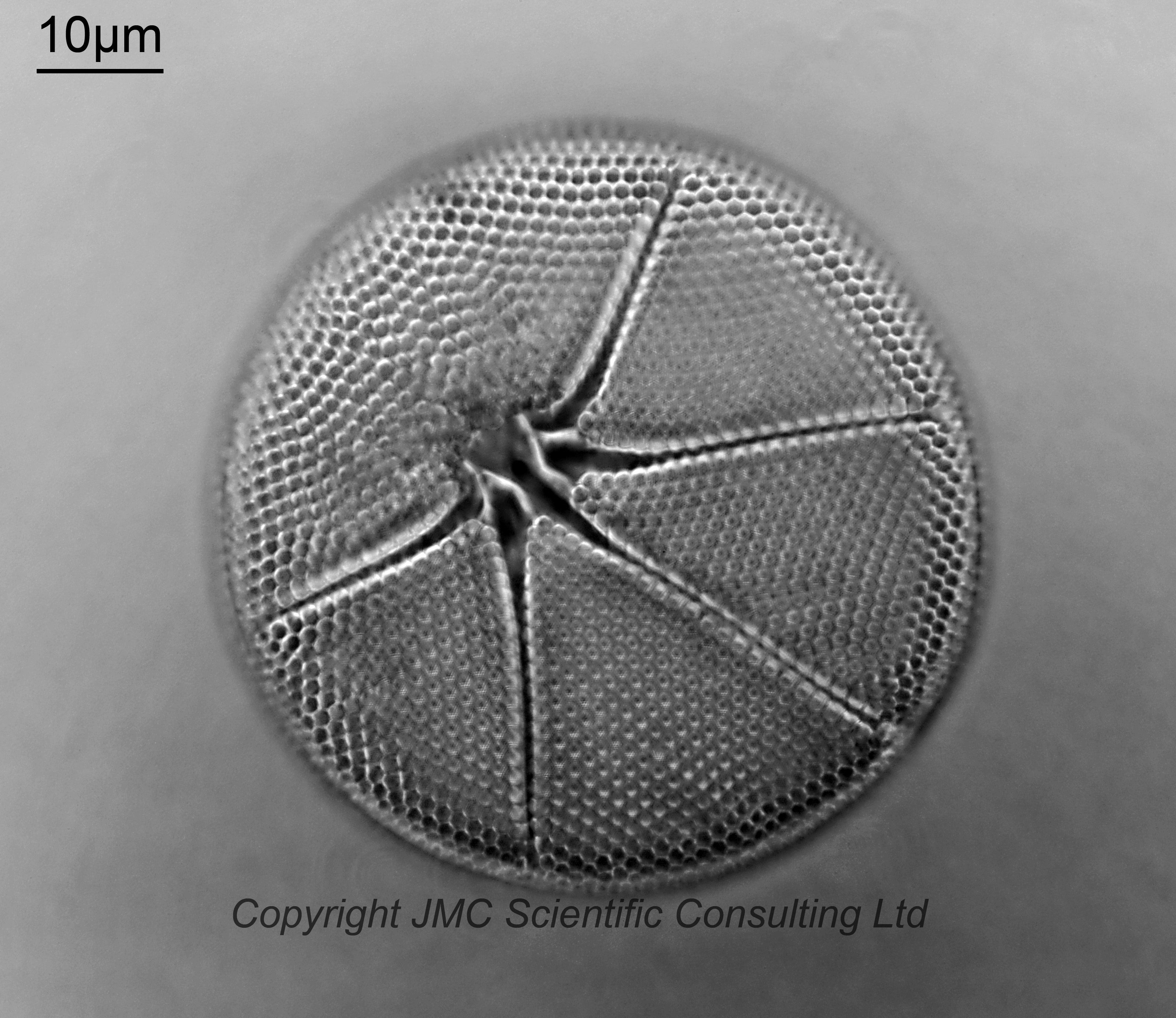
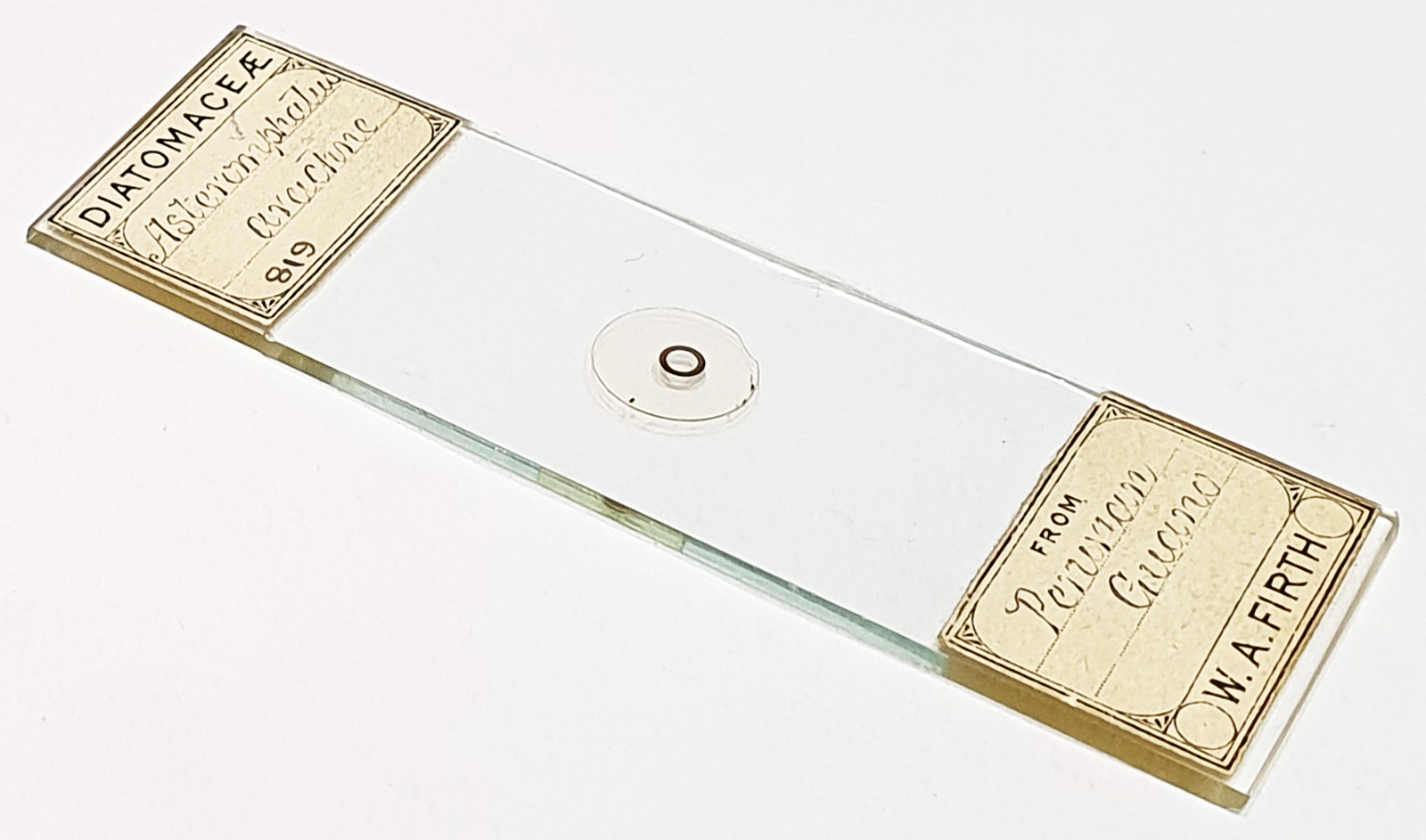
Two examples of Asteromphalus arachne from Peruvian guano. Accepted name for these is Spatangidium arachne Brébisson, 1857. Prepared by WA Firth. Olympus BHB microscope. Main image of the pair of them using 450nm LED light. 63x Leitz Pl Apo 1.4 objective, oil immersion. Olympus Aplanat Achromat condenser, oil immersion, oblique lighting. 2.5x Nikon CF PL photoeyepiece. Monochrome converted Nikon d850 camera. 39 images stacked in Zerene (Pmax).
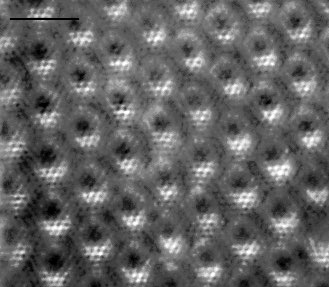
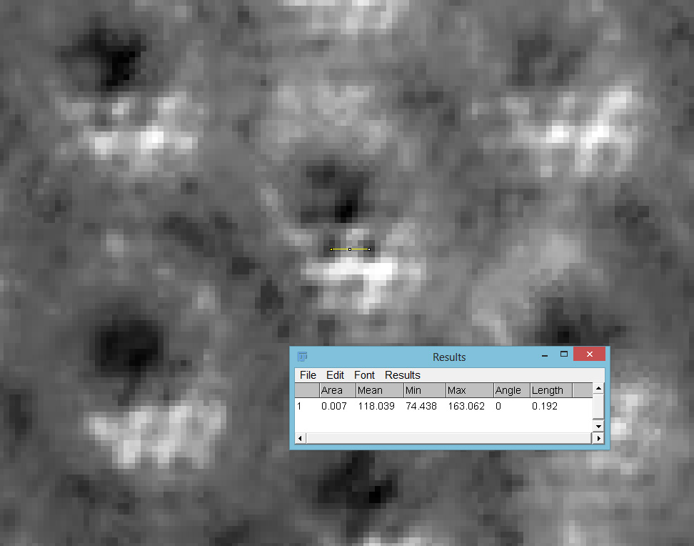
The entry for Asteromphalus arachne in the Diatom New Taxon File at the Academy of Natural Sciences, Philadelphia, shows some nice SEM images which show a hole pattern within their areolae. I wanted to try and visualize this but as it would be of the order of 200nm based on those images, I tried using 365nm light and a Reichert Neo 1.42/1.28 dark ground condenser, which gave circular oblique lighting (COL) with the 63x Leitz Pl Apo NA 1.40 objective. A single frame showed some nice dot patterns, and putting the image into ImageJ gave a dot spacing of 192nm. I fully accept that there will some error here, as the measurement in ImageJ depends on where I measured to and from, and with features this small, the precise locations of those ‘start’ and ‘stop’ points will be important. However that is in keeping with the spacings seen in the SEM images, so I am confident that what I was seeing weren’t just artifacts.