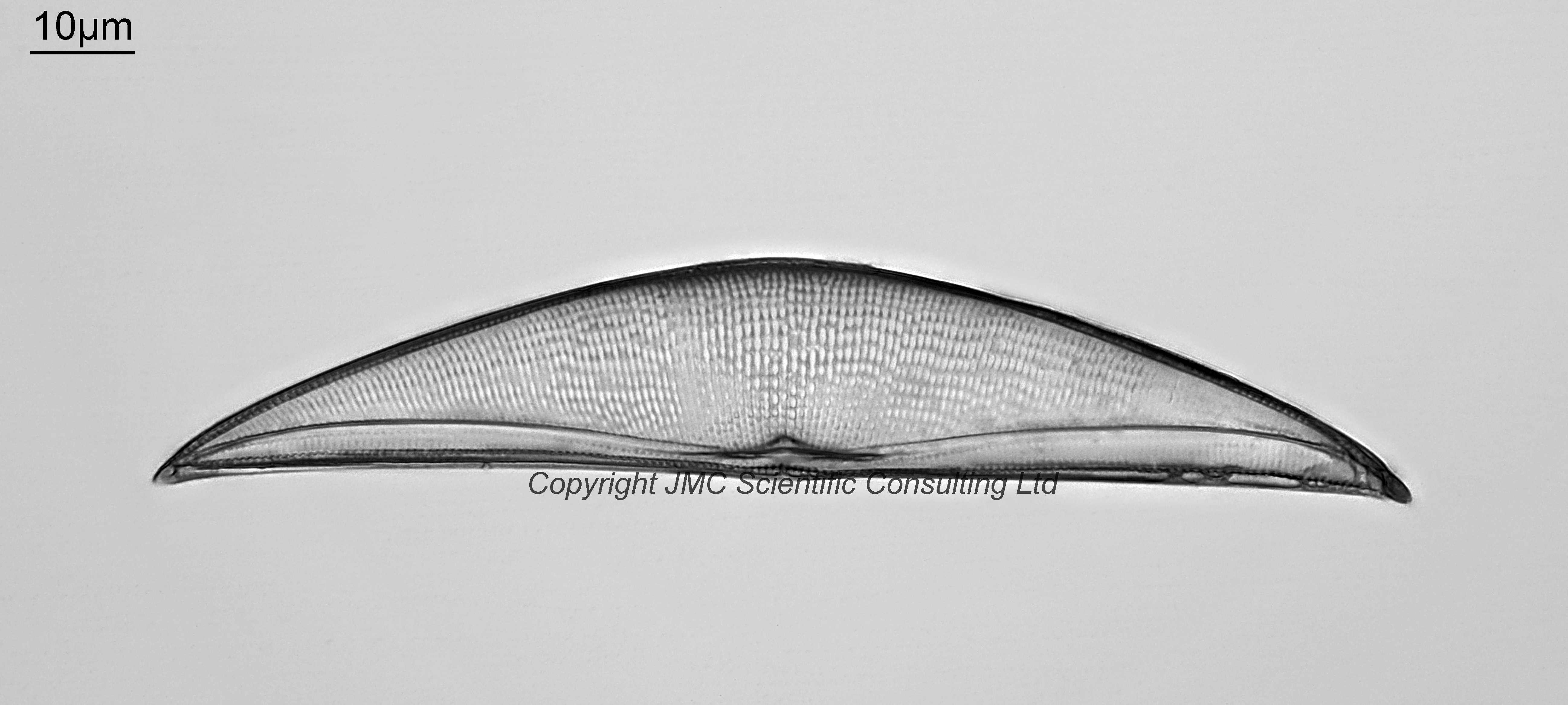
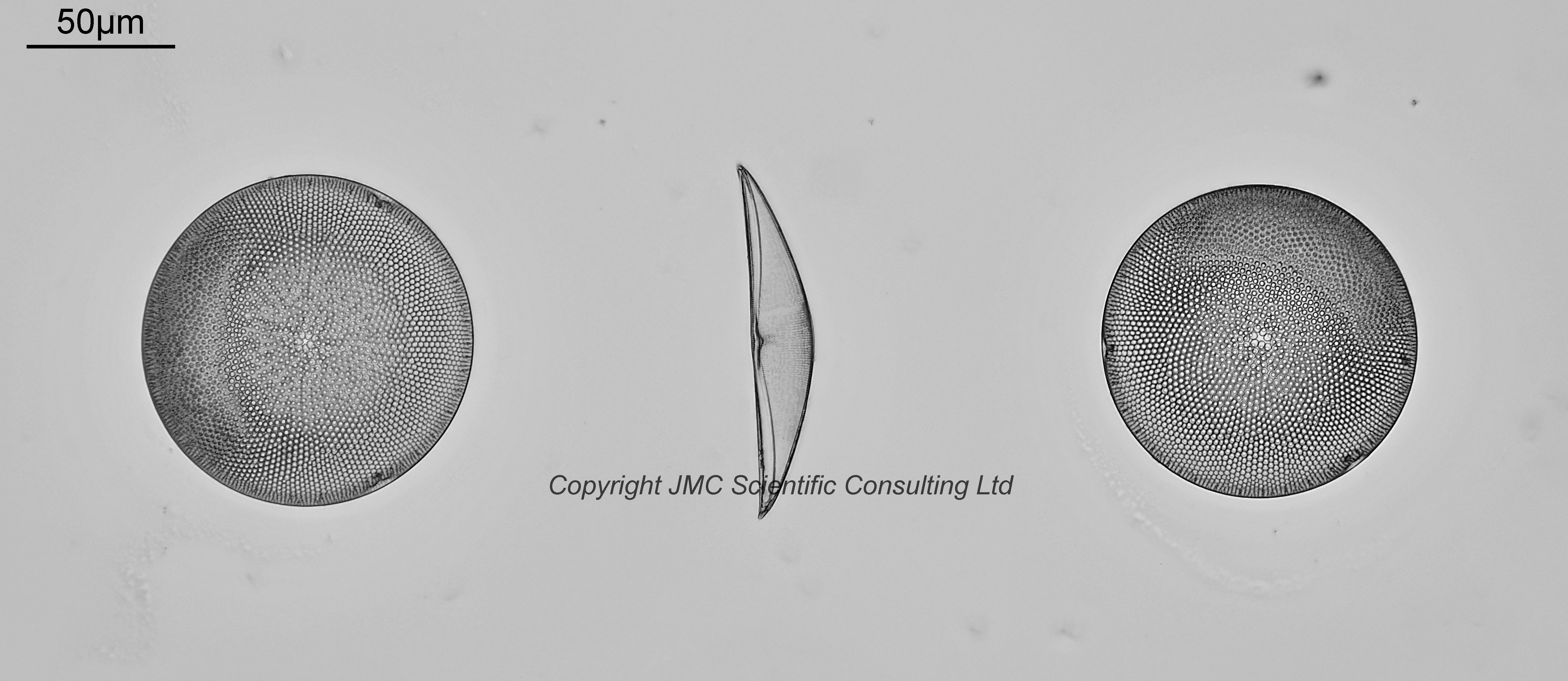
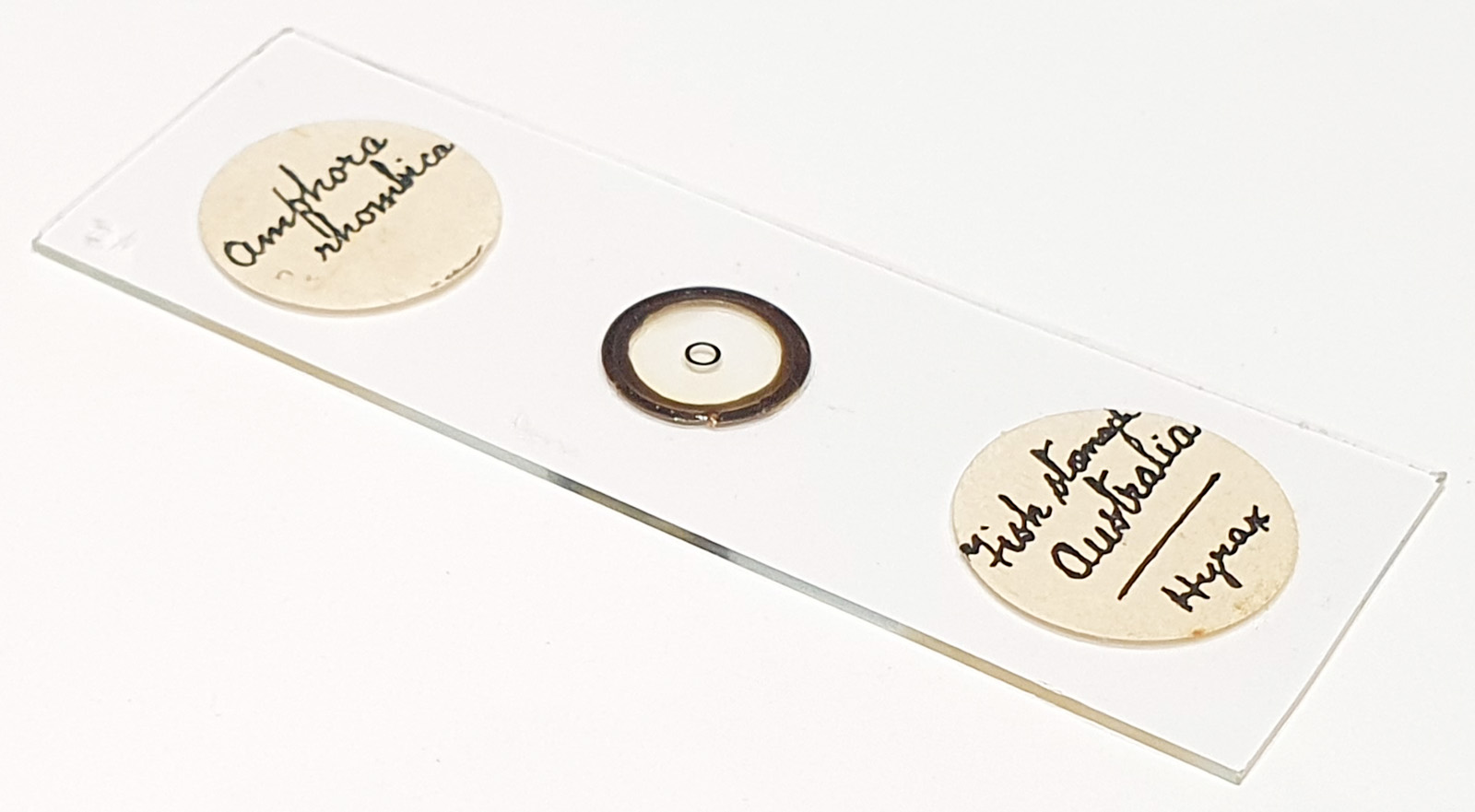
Amphora rhombica from fish stomach, Australia. Single example on the slide in between two circular diatoms. Prepared by JA Long, and mounted in Hyrax. Olympus BHB microscope using 390nm LED light. 63x Leitz Pl Apo NA 1.40 objective, oil immersion. Olympus Aplanat Achromat condenser, oil immersion, slightly oblique lighting. 2.5x Nikon CF PL photoeyepiece. Monochrome converted Nikon d850 camera. 17 images stacked in Zerene (Pmax). This was quite a low contrast diatom even with oblique lighting. It appears to be a ventral view (the inside of the diatom, rather than the convex outside).
I dug into the naming of this diatom when looking for examples to compare the image with, and came across a very good paper on the subject – Joshua G. Stepanek & J. Patrick Kociolek (2016) Re-examination of Mereschkowsky’s genus Tetramphora (Bacillariophyta) and its separation from Amphora,
Diatom Research, 31:2, 123-148, DOI: 10.1080/0269249X.2016.1183344. In this A. rhombica has two possible renamings – A. rhombica (Kitton in Schmidt) becomes Tetramphora rhombica (Kitton in Schmidt), and A. rhombica var. intermedia becomes Tetramphora intermedia (Cleve) Stepanek & Kociolek. Which one is this one? Good question. It’s length, tip to tip is 120µm. This puts it as small for T. rhombica – 130-260µm, but big for T. intermedia – 80-105µm. Likewise the striae separation for this one (13.1 striae per 10µm), puts it between the two. However in Cleve, 1895, Synopsis of naviculoid diatoms. II. Kongliga Svenska-Vetenskaps Akademiens Handlingar 27: p. 127, the striae count on the ventral side is given as 11-13 per 10µm for A. rhombica (Kitton), and 16 per 10µm for var. intermedia. Based on this I would say this one is A. rhombica (Kitton in Schmidt), newer name Tetramphora rhombica (Kitton in Schmidt), but a small example of it. I got there in the end.