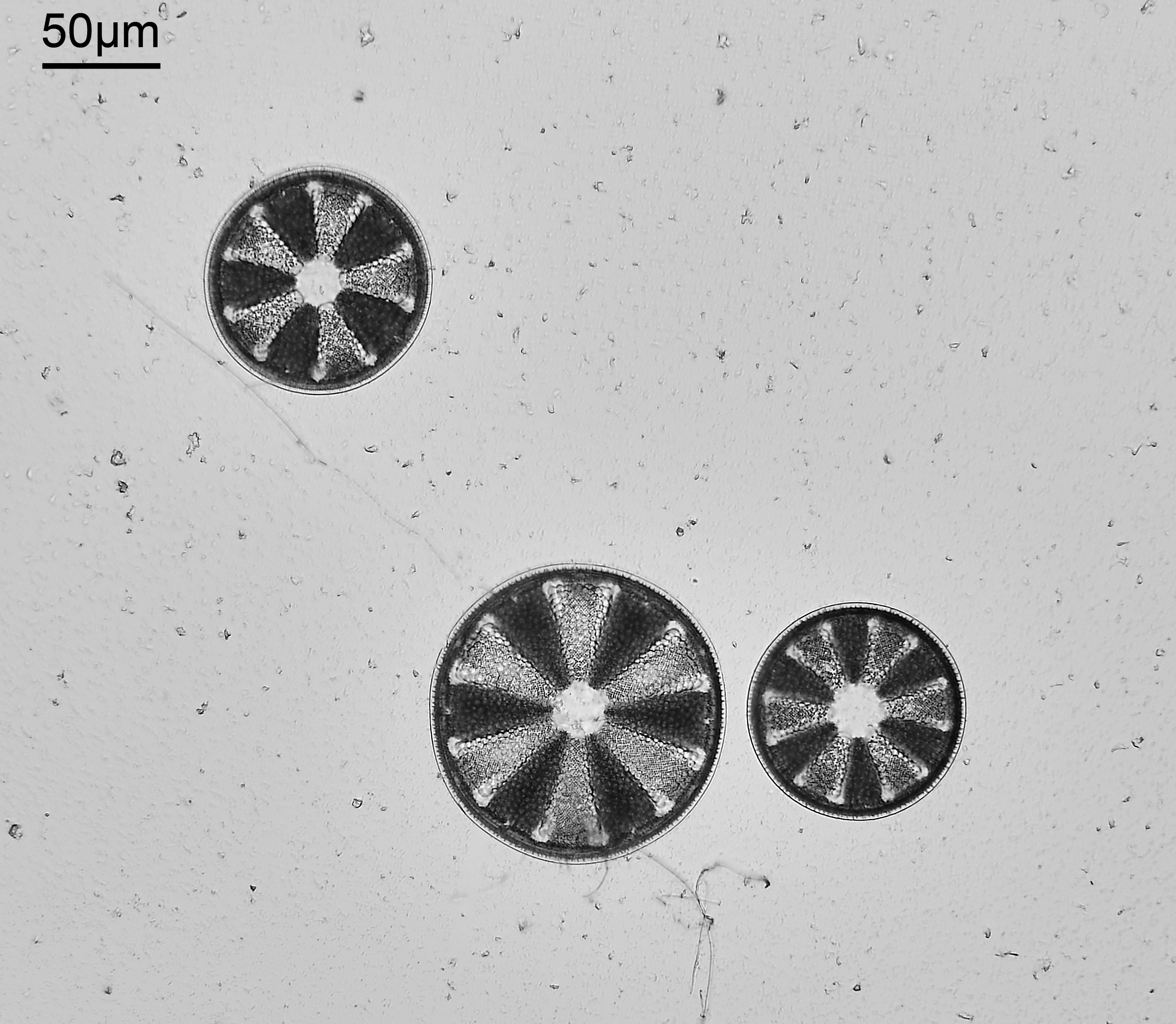
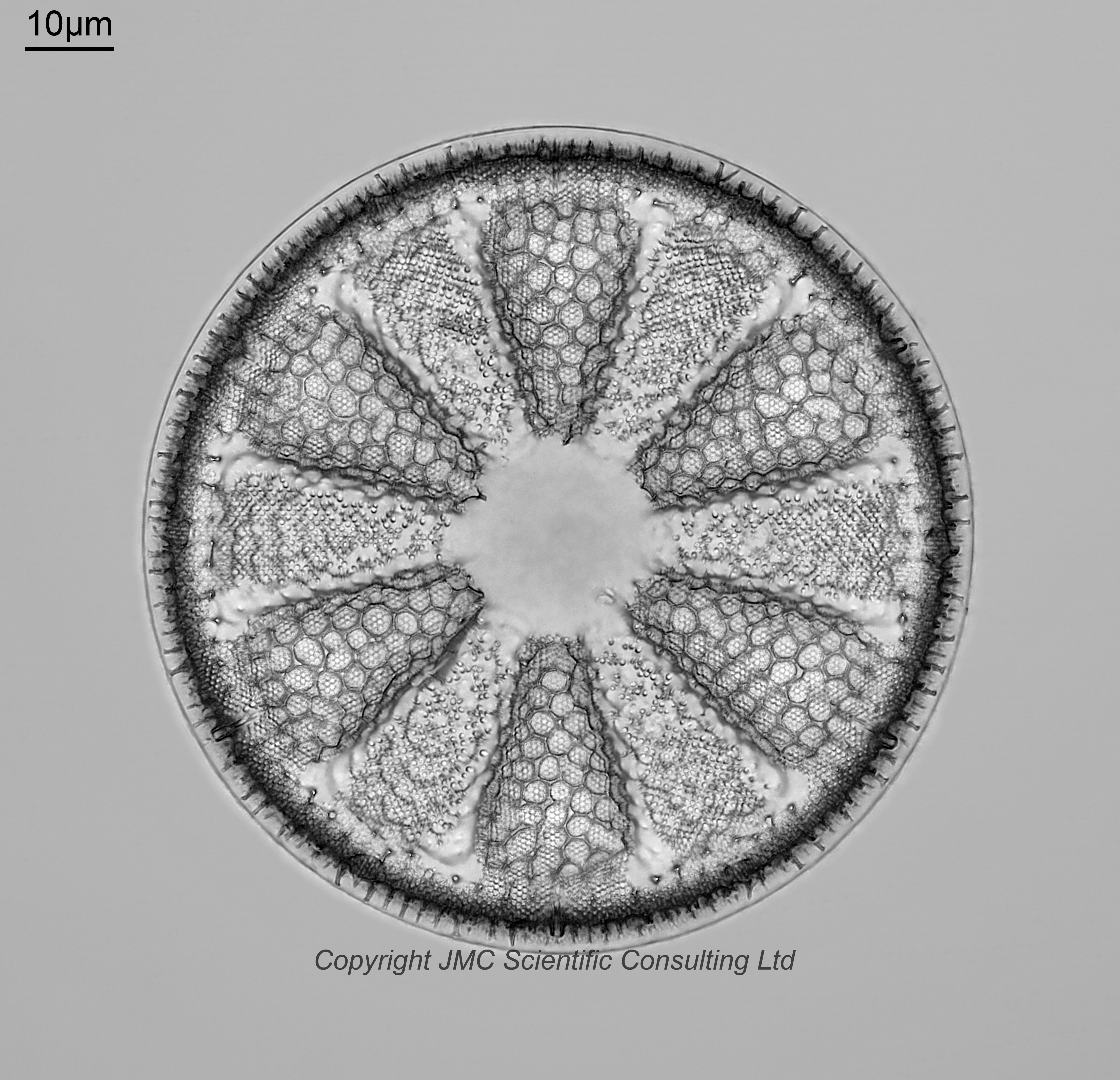
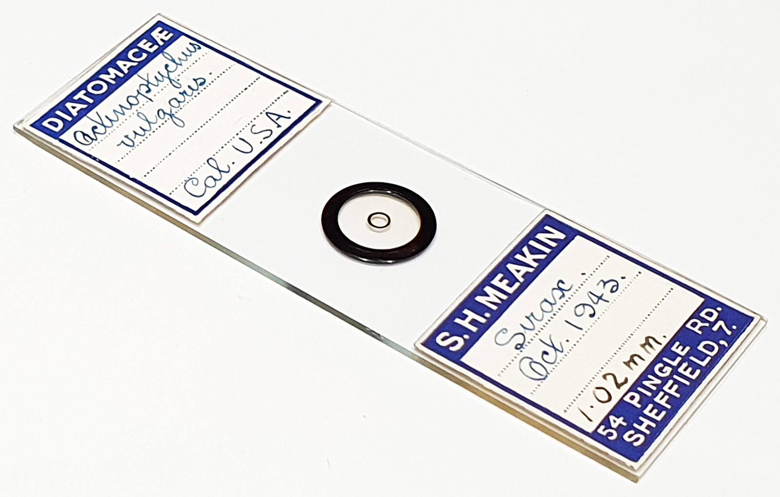
A loose arrangement of three Actinoptychus vulgaris from California, USA. Prepared by Samuel Henry Meakin. Mounted in Styrax and dated October 1943. Olympus BHB microscope using 450nm LED light. 63x Leitz Pl Apo 1.4 objective, oil immersion. Olympus Aplanat Achromat condenser, oil immersion, brightfield lighting. 2.5x Nikon CF PL photoeyepiece. Monochrome converted Nikon d850 camera. 28 images stacked in Zerene (Pmax).
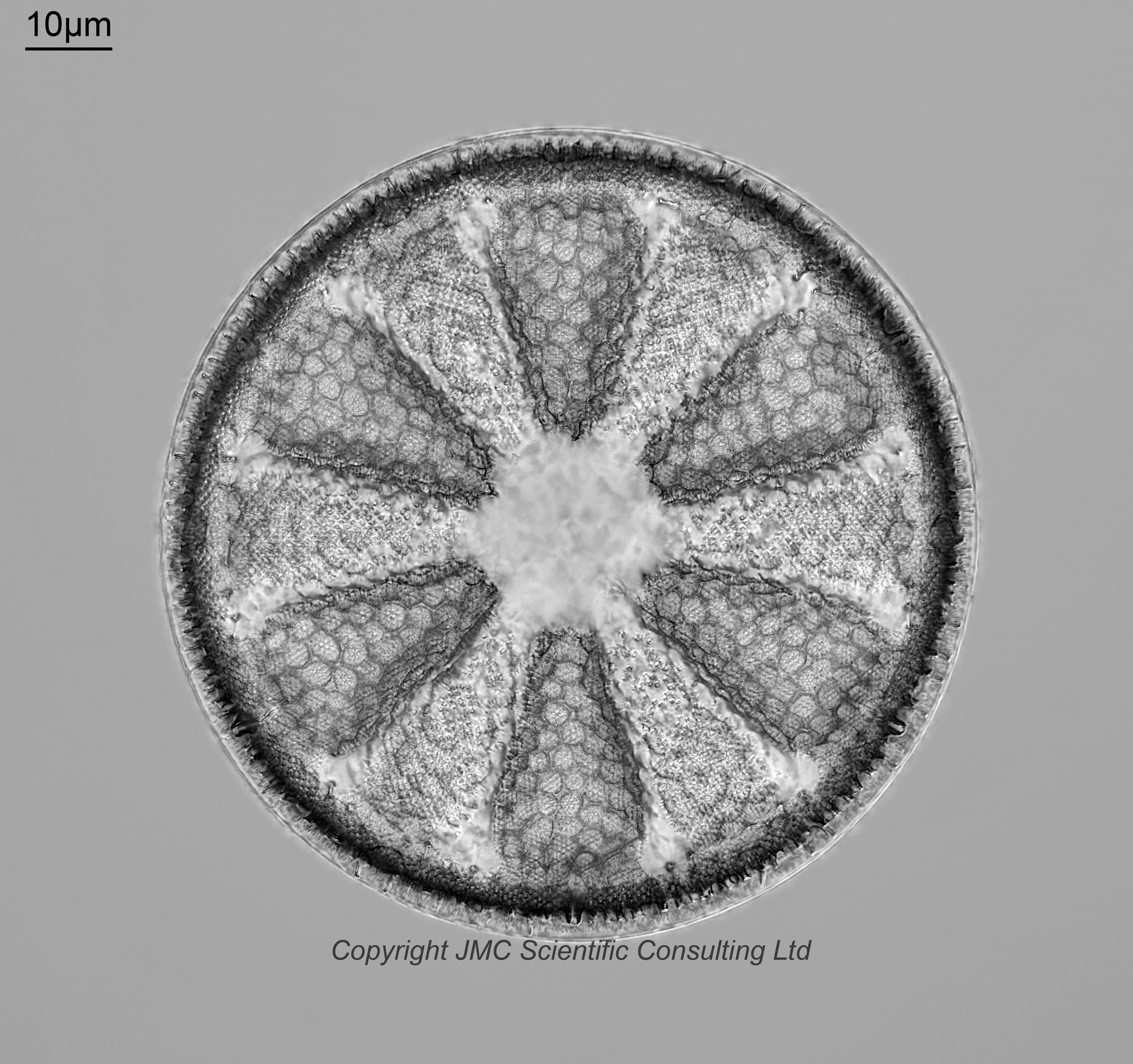
Those of your that know my work know my interest in UV microscopy. Not fluorescence work, but using UV light for transmission microscopy and capturing UV photos, due to the potential it has for improved resolution. Sirax is a mountant which has pretty good UV transmission at 365nm, so I decided to also image the same diatom with 365nm LED light. I did use a standard Olympus Abbe condenser, as the Aplanat Achromat blocks UV, but the other optics were kept the same. And, yes I did use the 63x Leitz Pl Apo objective for 365nm. Although well out side of it’s design wavelength it works quite well for 365nm imaging. The 365nm image was a bit disappointing and did not look as clear as the 450nm one. I think one of the issues here was that there was a lot of structure present in the mountant, and this became more obvious at 365nm vs 450nm. This structure can be seen in the 10x objective image, but obviously I have removed it from around the diatom in the high magnification images. Although removed from around the diatom it is still visible through the diatom as the images are captured for stacking. This in turn then obscured some of the details in the diatom during stacking as this ‘structure’ became part of the stacked image. Moral of this story, there is often a mismatch between theory and and experiment, and one should never assume the outcome of the experiment.