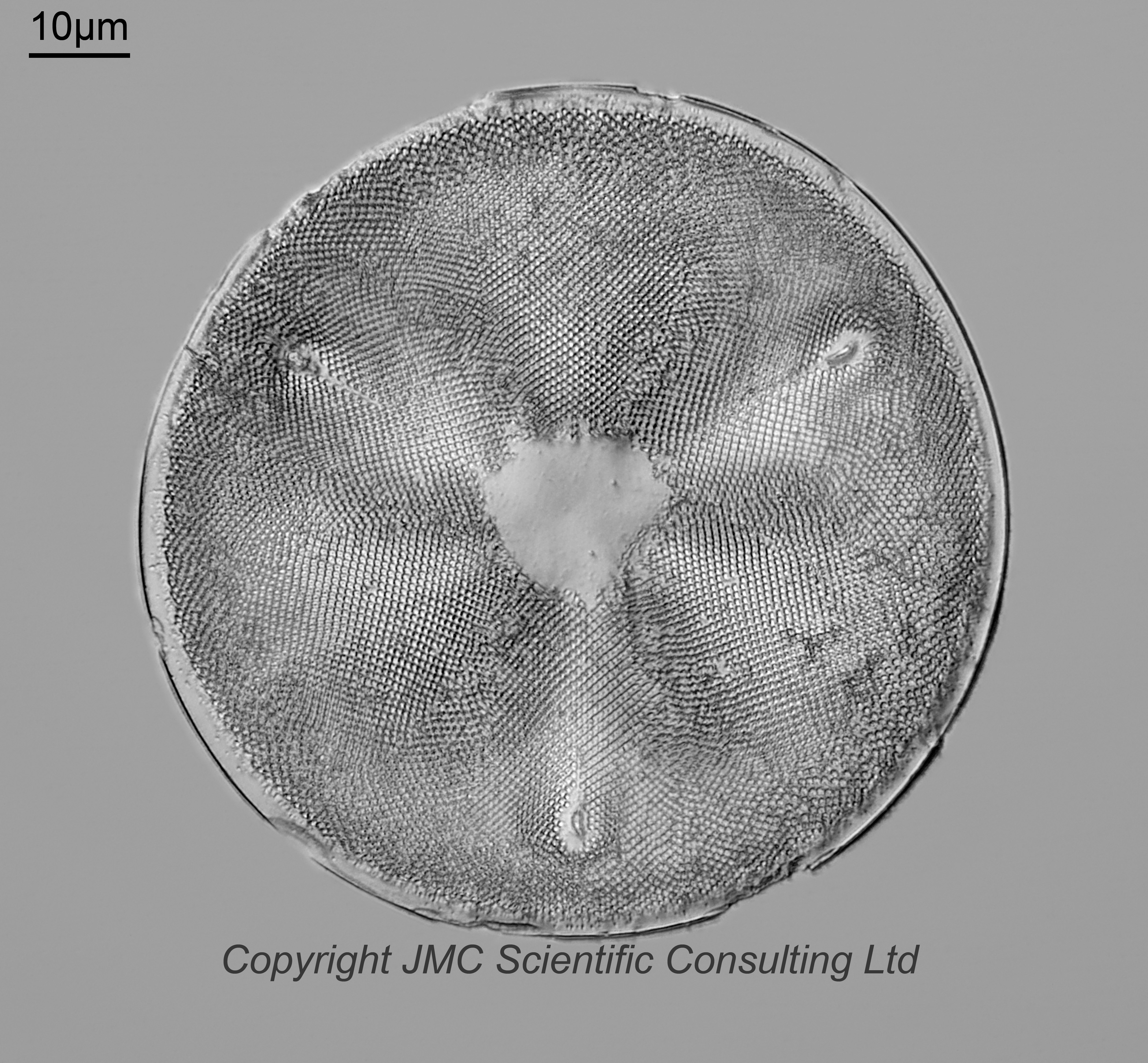
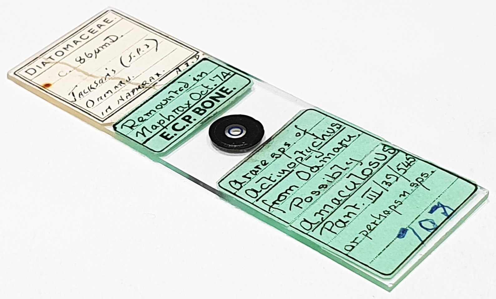
An interesting (and a little puzzling) slide of an Actinoptychus diatom from Jackson’s [Paddock], Oamaru, New Zealand. Single example on the slide. Mounted in Naphrax. 86µm diameter. Looks to have been remounted in Naphrax in October 1974 by ECP Bone. The label says “A rare species of Actinoptychus from Oamaru. Possible A. maculosus Pant III/39/545 or perhaps a new species”. I’ll come back to the name in a minute. Olympus BHB microscope using 450nm LED light. 63x Leitz Pl Apo 1.4 objective, oil immersion. Olympus Aplanat Achromat condenser, oil immersion, oblique lighting. 2.5x Nikon CF PL photoeyepiece. Monochrome converted Nikon d850 camera. 28 images stacked in Zerene (Pmax).
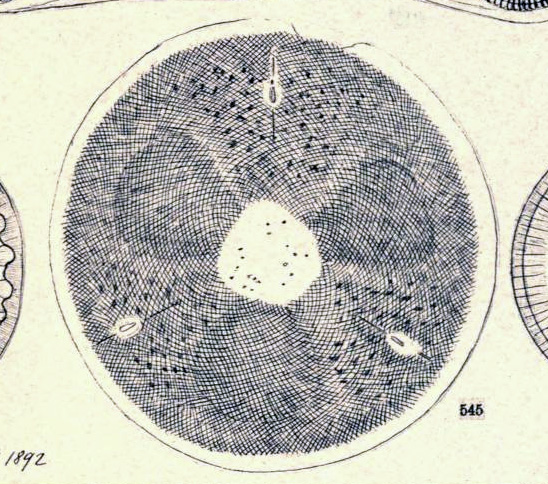
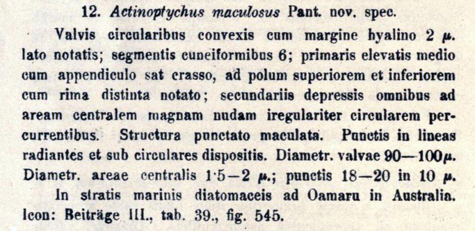
Back to the name. Actinoptychus maculosus Pantocsek, 1892. Pantocsek, J. (1892). Beiträge zur Kenntnis der Fossilen Bacillarien Ungarns. 3 Teile. Julius Platzko, Nagyy-Tapolcsany. Teil III, Susswasser Bacillarien. Anhang-analysen 15 neuer Depots von bulgarien, Japan, Mahern, Russland und Ungarn. Nagy-Tapolcsány, Buchdrucherei von Julius Platzko, 42 pls., available online at http://www.biodiversitylibrary.org/item/15525. Page 6, Plate 39, Figure 545. I’ve shown screenshots of the image for it, and the text description above. The translation of the text is given below;
“Circular valves convex with a hyaline margin 2 µm. wide; cuneiform segments 6; primary elevated in the middle with a fairly thick appendage, marked at the upper and inner poles with a distinct groove; secondaries depressed all running to a large bare irregularly circular central area. Structure punctate-spotted. Punctate arranged in radiating and subcircular lines. Valve diameter 90-100 µm. Central area diameter 1.5-2 µm. (?); punctate 18-20 in 10 µm.
In diatomaceous marine strata at Oamaru in Australia [sic]. Icon: Contributions (?) III., Plate 39., Figure 545.”
The appearance of A. maculosus is pretty close to this one. However there are some errors in Pantocsek’s text. Oamaru is in New Zealand, not Australia. Also, the central area looks to be more like 15-20µm across not 1.5-2µm. I cannot find other images of A. maculosus to compare with.
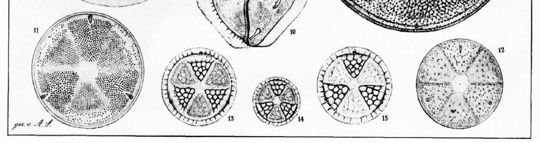
Looking at other sources, it is similar to Actinoptychus simbirskianus (Schmidt’s Atlas, Plate 29, Figure 11). However to me it is more like Actinoptychus seductilis (Schmidt’s Atlas Plate 29, Figure 12) based on the placement of the three processes in from the edge. However does look like A. simbirskianus from Oamaru Diatoms website. There therefore seems to be a mismatch between Oamaru Diatoms website and Schmidt’s Atlas based on the position of the processes. BUT, it looks like A. simbirskianus from the image in Desikachary and Sreelatha’s Oamaru book though. However I also cannot rule out Actinoptychus trilunatus from Desikachary and Sreelatha’s (would need more/better pictures to compare against though).
Also, Desikachary and Sreelatha’s Oamaru book does not mention A. maculosus.
Overall a puzzling slide. Actinoptychus, yes. Which species, not sure.