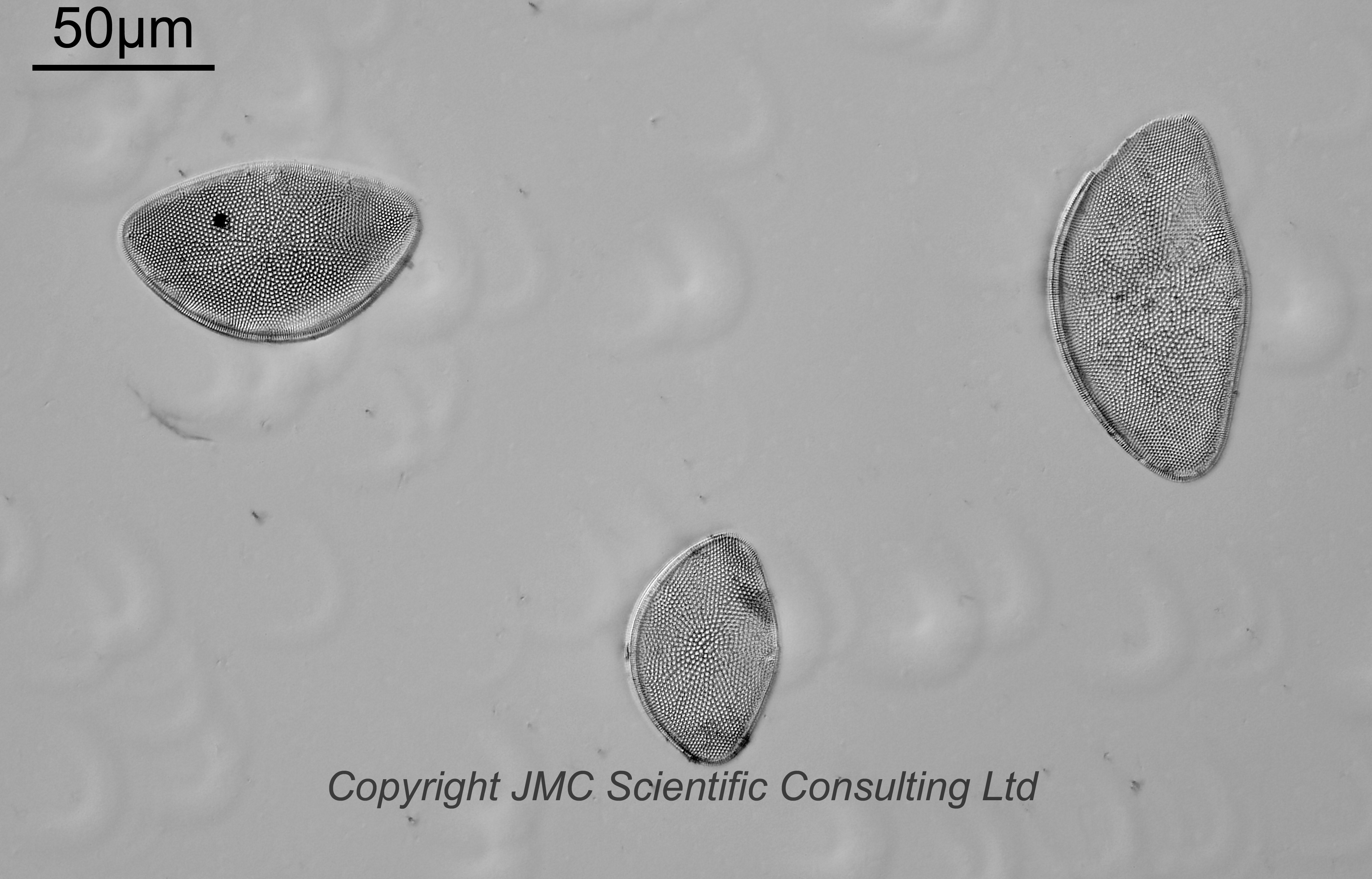
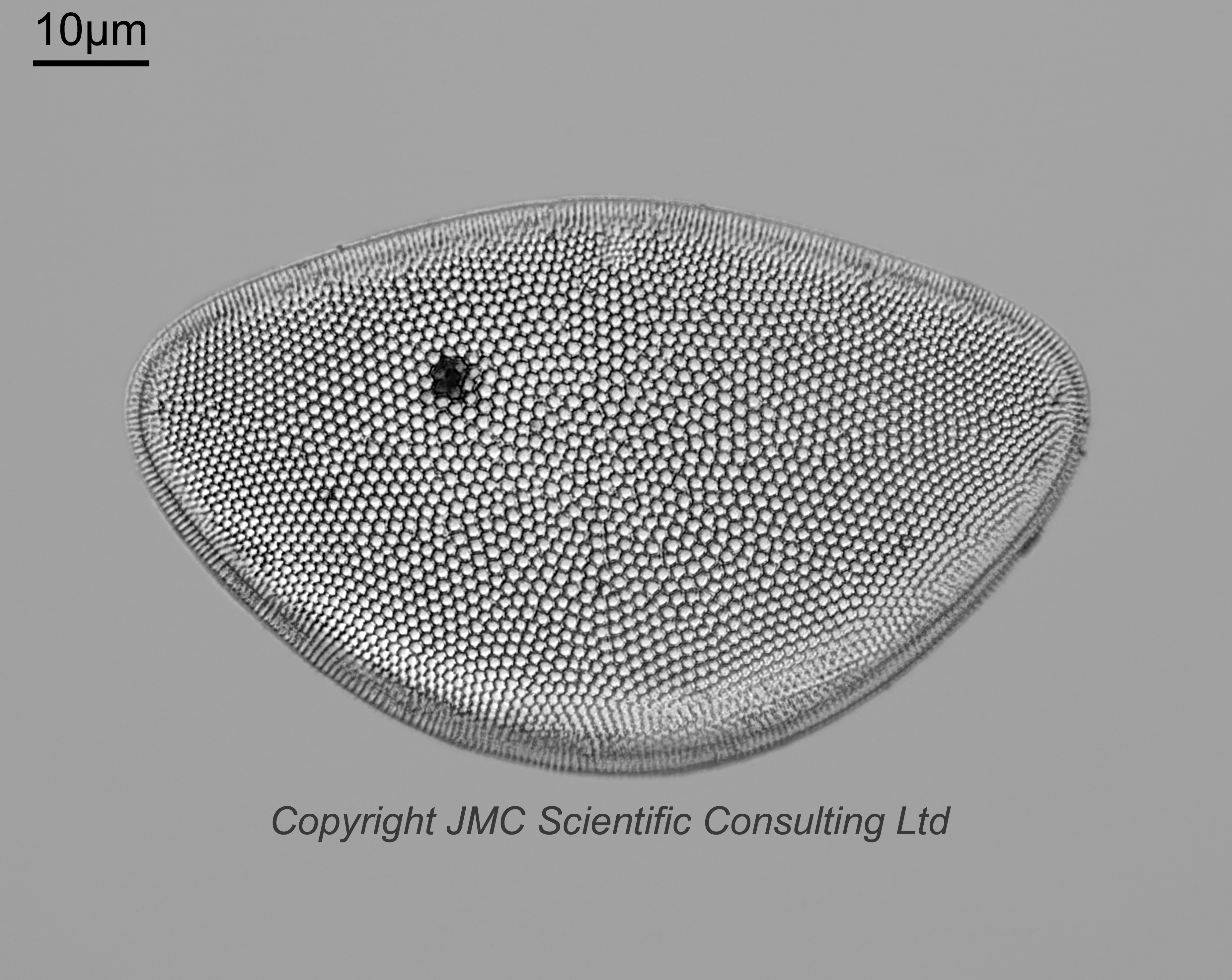
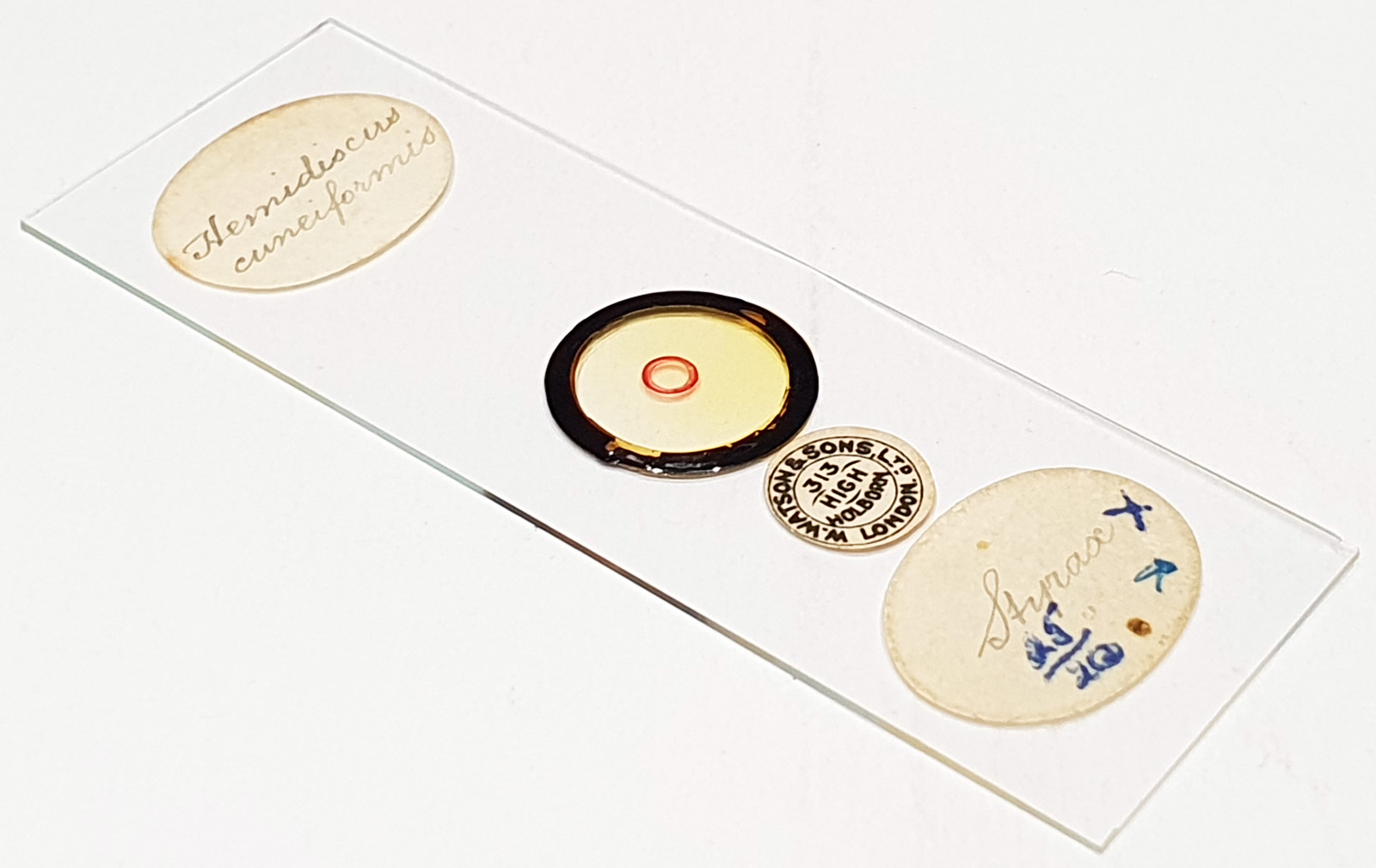
Three examples of Hemidiscus cuneiformis prepared by Watson and Sons Ltd. Some ‘bubbles/lenses’ in the mountant. Olympus BHB microscope using 450nm LED light. As well as the group, I imaged one using the 63x Leitz Pl Apo 1.4 objective, oil immersion. Olympus Aplanat Achromat condenser, oil immersion, oblique lighting. 2.5x Nikon CF PL photoeyepiece. Monochrome converted Nikon d850 camera. 45 images stacked in Zerene (Pmax). Dipping down away from the coverslip towards the top edge.
Hemidiscus cuneiformis Wallich 1860. Published in: Wallich, G.C. (1860). On the siliceous organisms found in the digestive cavities of the Salpae, and their relation to the flint nodules of the Chalk Formation. Transactions of the Microscopical Society of London, New Series 8: 36-55, pl. II. Page 42, Plate II [2], Figures 3,4. This name is currently regarded as a synonym of Actinocyclus cuneiformis (Wallich) F.Gómez, Lu Wang & Senjie Lin 2017. Published in: Gómez, F., Wang, L., Hernández-Becerril, D.U., Lisunova, Y.O., Lopes, R.M. & Lin, S. (2017). Molecular phylogeny suggests transfer of Hemidiscus into Actinocyclus (Coscinodiscales, Coscinodiscophyceae). Diatom Research 32(1): 21-28. Page 26, [figures. 1–23].