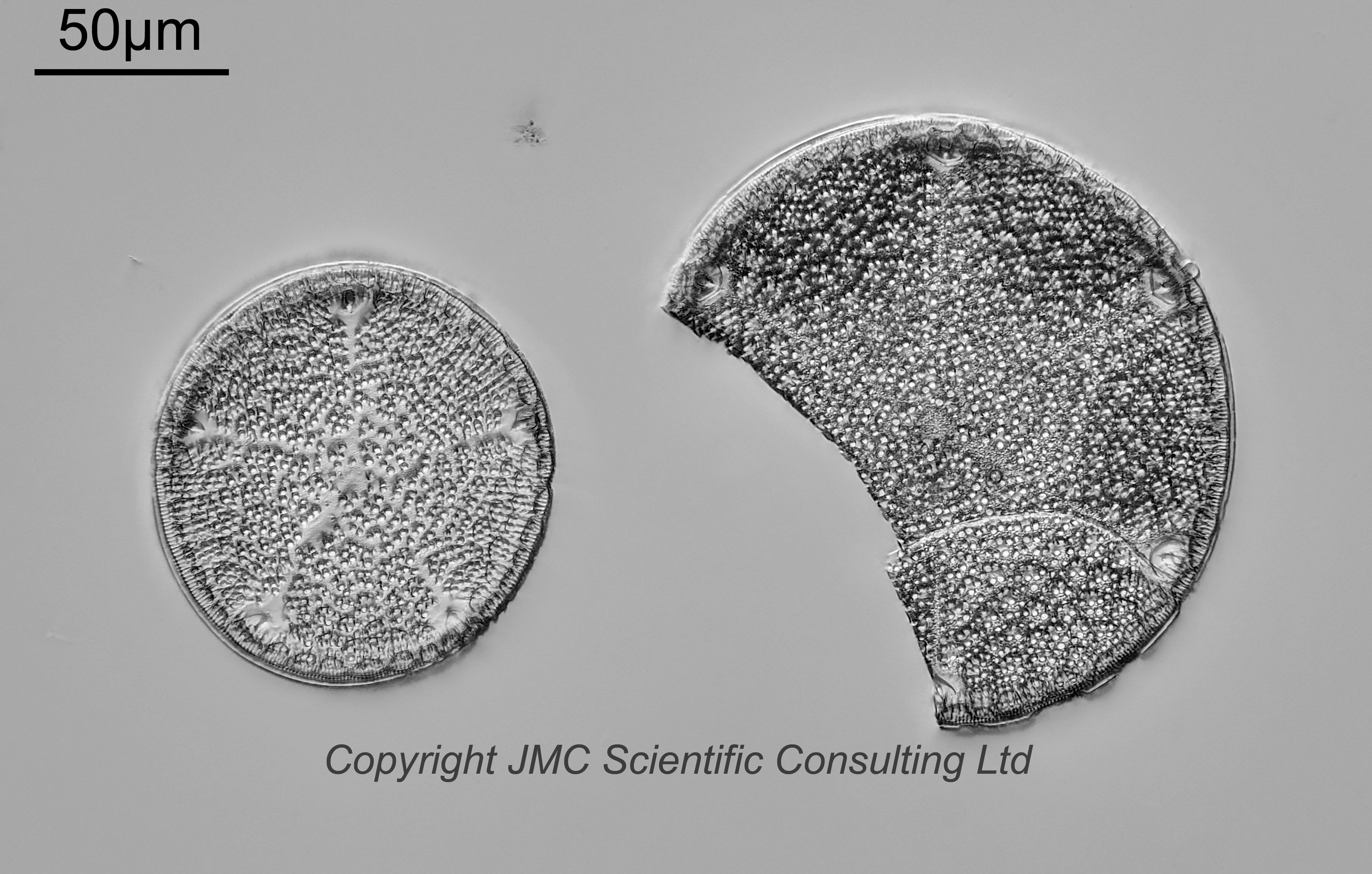
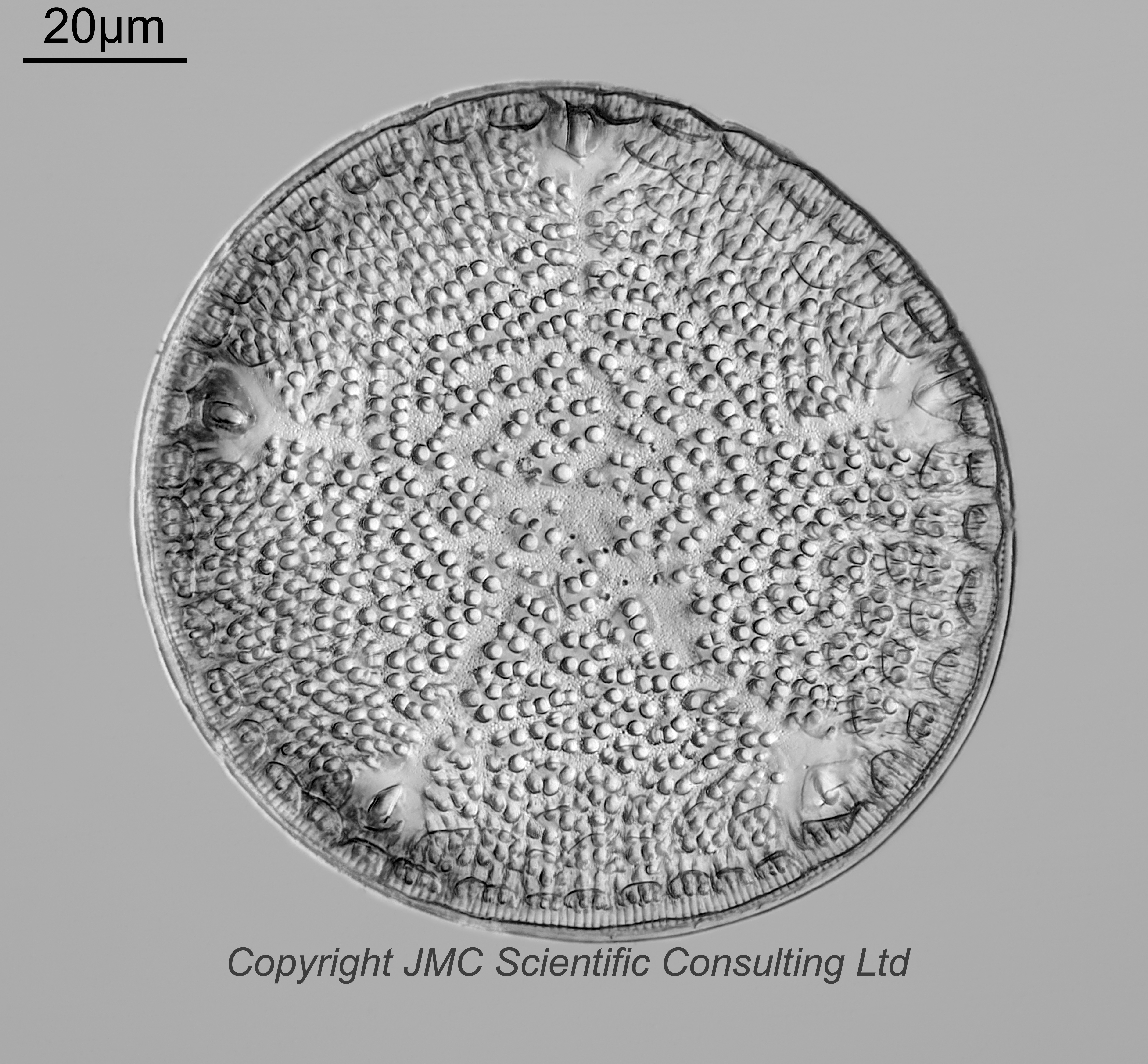

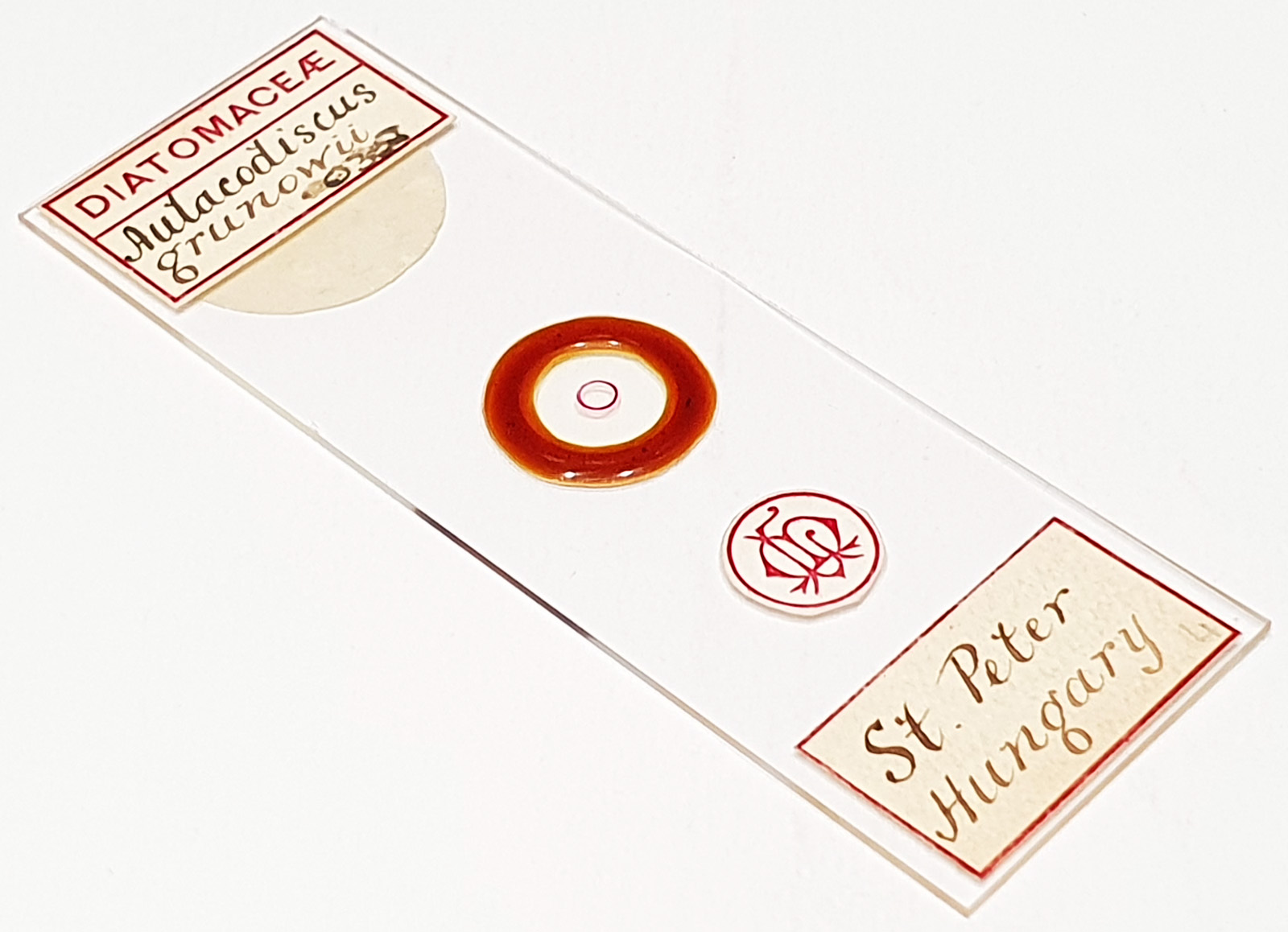
A slide with two examples of Aulacodiscus grunowii from St Peter, Hungary. Prepared by Laurence Miles. Olympus BHB microscope using 450nm LED light. As well as the arrangement I also imaged the smaller one with 5 processes using the 63x Leitz Pl Apo NA 1.4 objective, oil immersion. Olympus Aplanat Achromat condenser, oil immersion, oblique lighting. 2.5x Nikon CF PL photoeyepiece. Monochrome converted Nikon d850 camera. 67 images stacked in Zerene (Pmax). The smaller one had a diameter of 105-110µm diameter. Stacking has lost the top layer of features (the ‘reticulations’?), so I am including a partial stack to show some of these more subtle features.
Aulacodiscus grunowii Cleve 1885. Published in: Cleve, P.T. (1885). On some fossil marine diatoms found in the Moravian “Tegel” from Augarten, near Brünn. Journal of the Quekett Microscopical Club, Series 2 2(13): 165–177, plates 12–13. Page 171, Plate XII [12], Figure 8 (as ‘Grunowii‘).
These two have a different look to each other. The smaller one with 5 processes is more dome shaped, while the larger partial one is more of a truncated cone. I have examples of A. grunowii on this site with both types of morphology. Original description by Cleve says 6-12 processes for thsi species, and a reticulated layer below the granular surface for A. grunowii, and a large diameter of 100-300µm. The smaller one has only 5 processes and is at the bottom end of the size scale given by Cleve.