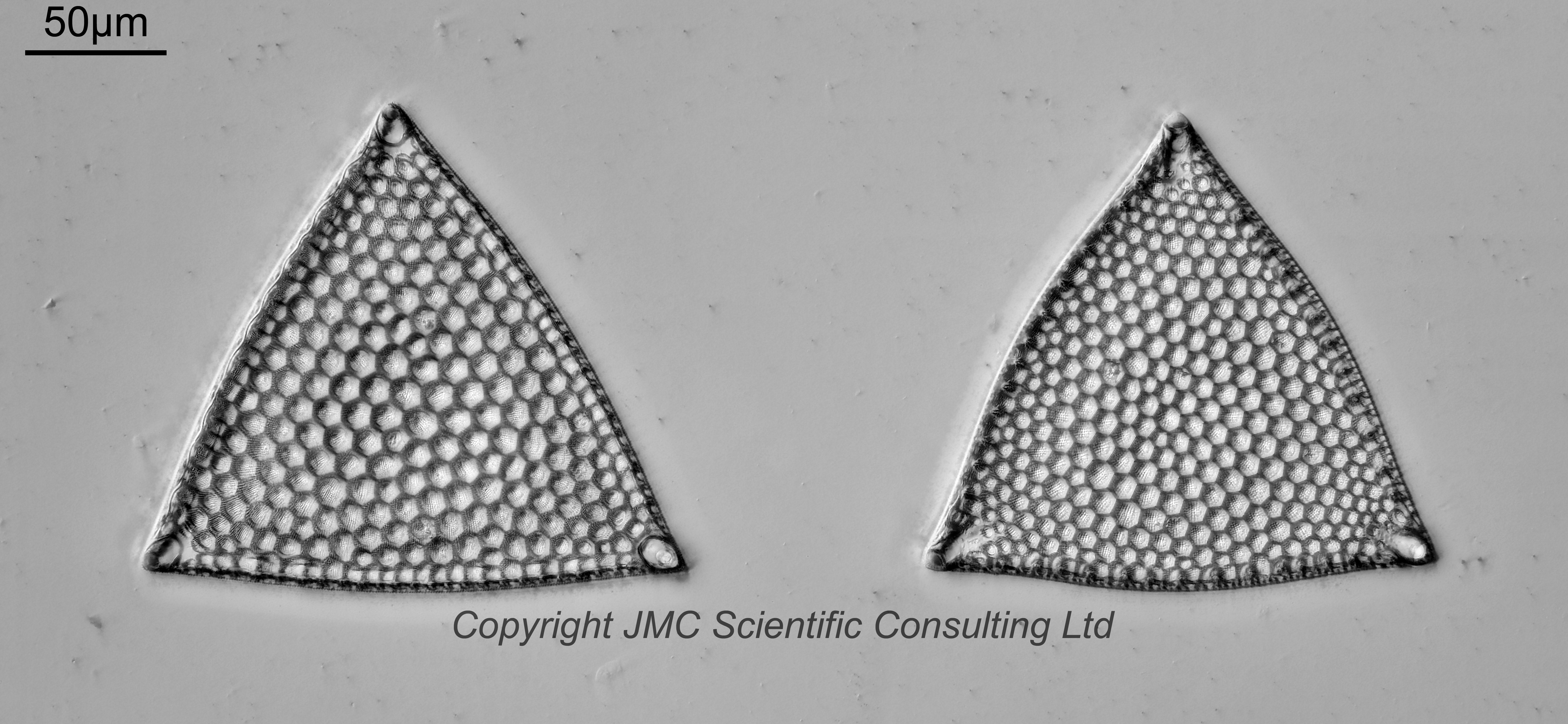
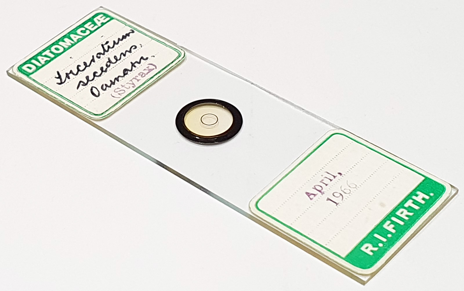
Slide labelled as Triceratium secedens (however I don’t think either of these are that, more later) from Oamaru, New Zealand. Mounted in Styrax and dated April 1966. Prepared by RI Firth. Olympus BHB microscope using 450nm LED light. As well as the arrangement both were imaged individually using a 63x Leitz Pl Apo 1.4 objective, oil immersion. Olympus Aplanat Achromat condenser, oil immersion, oblique lighting. 2.5x Nikon CF PL photoeyepiece. Monochrome converted Nikon d850 camera. Image stacks prepared in Zerene (Pmax).
Back to the names. Firstly these look to be two different species to me. Neither of these look like T. secedens (again, this is to me). Here’s my thoughts as to what they are.
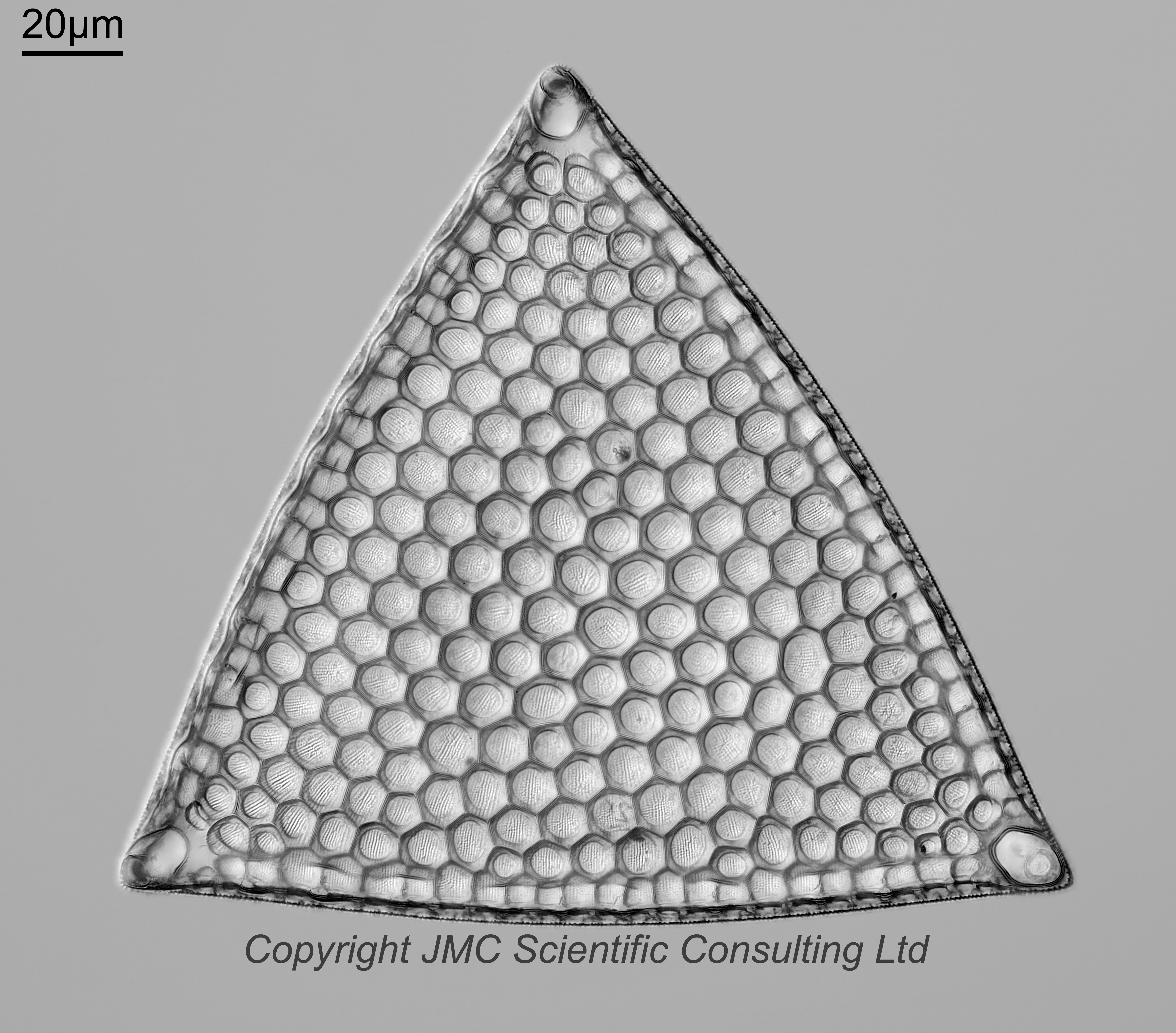
The left one. 52 images stacked. Triceratium favus var. maximum Grunow 1882:. Published in: Van Heurck, H. (1882). Synopsis des Diatomées de Belgique. Atlas. pp. pls LXXVIII-CXXXII [78-132 and supplementary pls 22 bis, 82bis, 83bis, 83ter, 95bis], tables genres. Anvers: Ducaju et Cie. Plate CVII [107], Figure 5 (as ‘maxima‘). See also Desikachary, T.V. & Sreelatha, P.M. (1989). Oamaru diatoms. Bibliotheca Diatomologica 19: [1]-330, including 145 plates. Pages 255-256 (as ‘maxima‘), Plate 121, Figure 4.
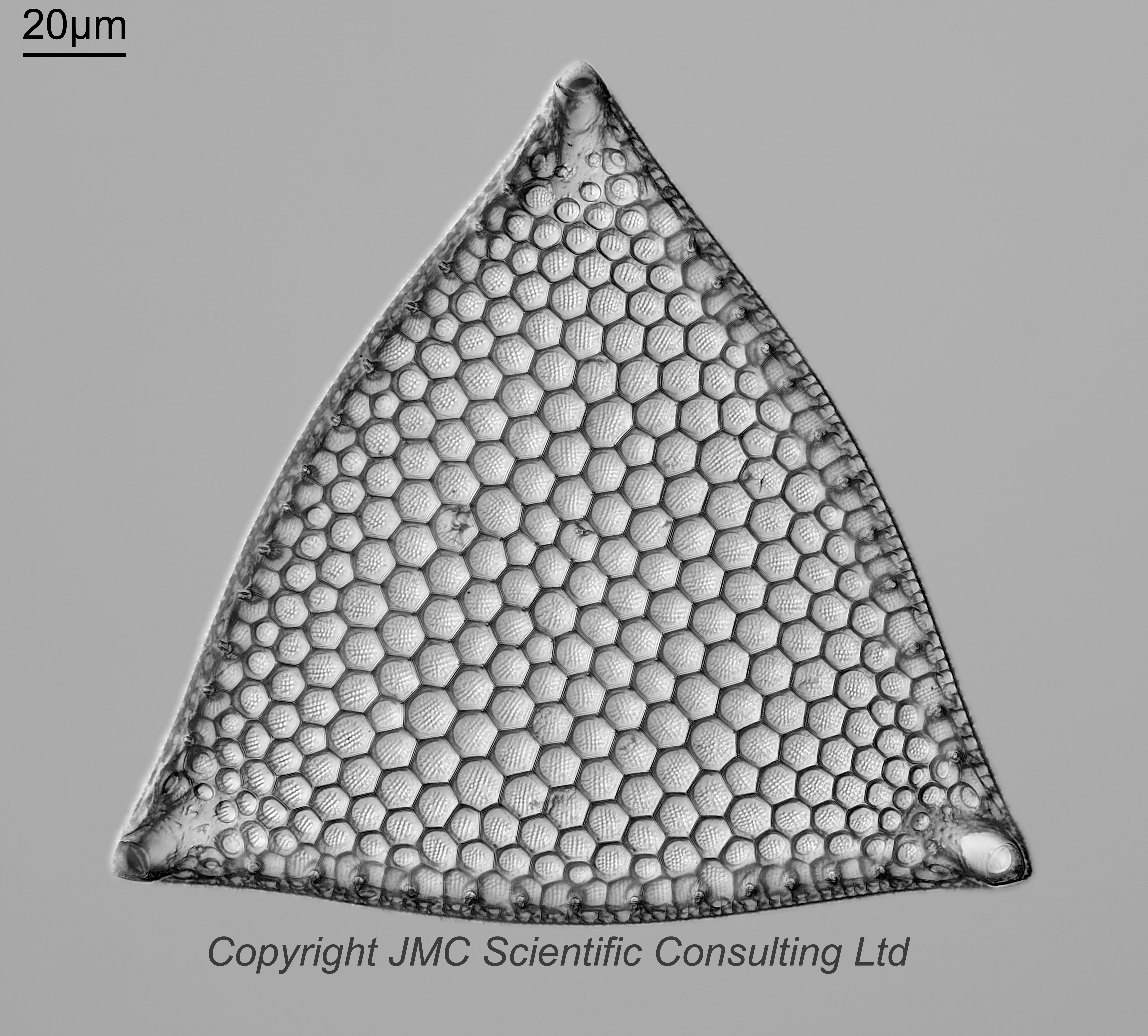
The right one. 58 images stacked. Spikes around the edge. Triceratium broeckii Leuduger-Fortmorel 1879. Published in: Leuduger-Fortmorel, G. (1879). Catalogue des Diatomées de l’Ile Ceylan. pp. 1-73, 9 pls [I-IX]. Saint-Brieuc: Librairie Francisque Guyon, Impriméur. Page 61, Plate 6, Figure 63. See also Desikachary, T.V. & Sreelatha, P.M. (1989). Oamaru diatoms. Bibliotheca Diatomologica 19: [1]-330, including 145 plates. Page 249, Plate 116, Figures 6,9.
I will leave both of these with a question mark for now though.