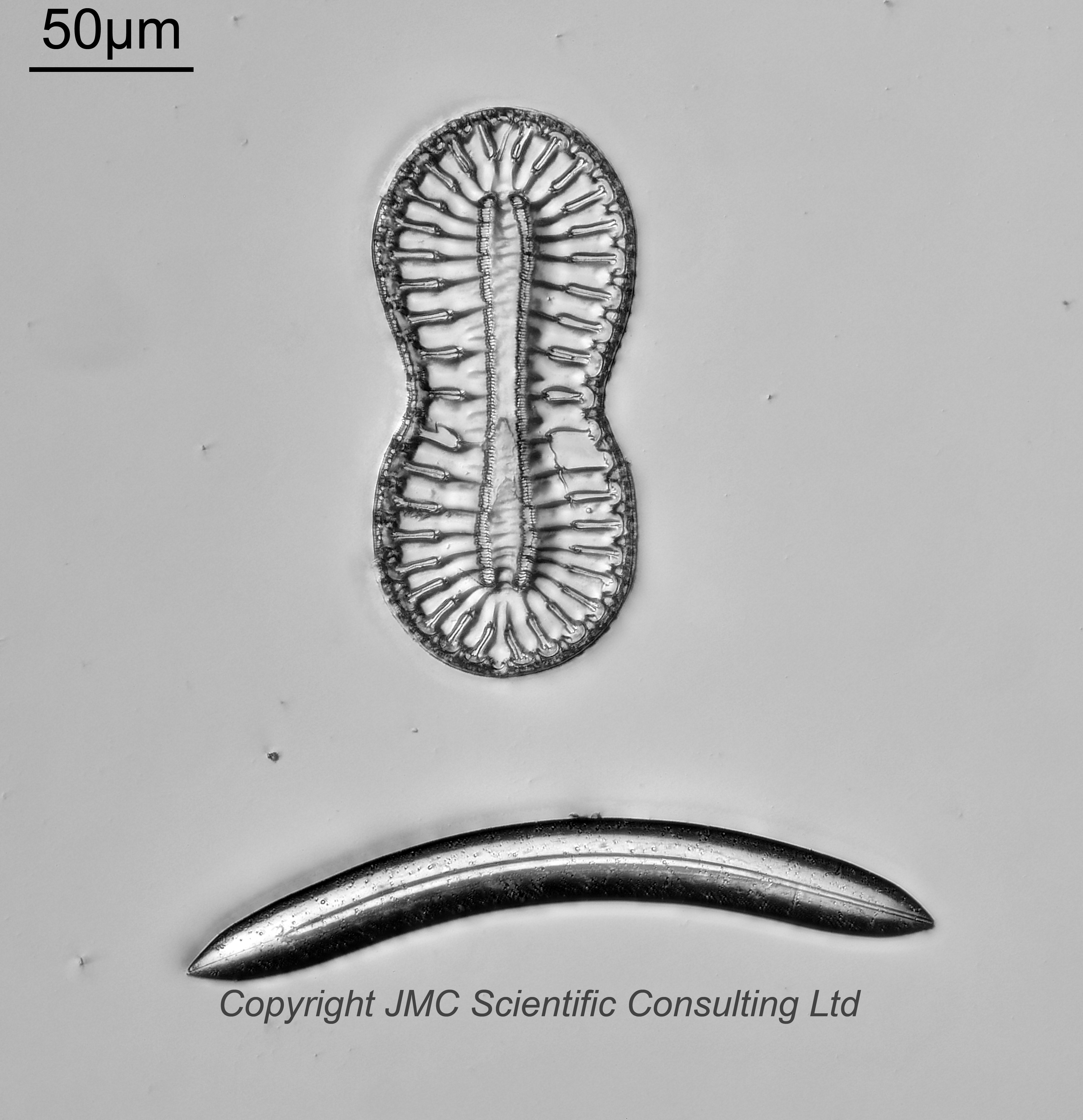
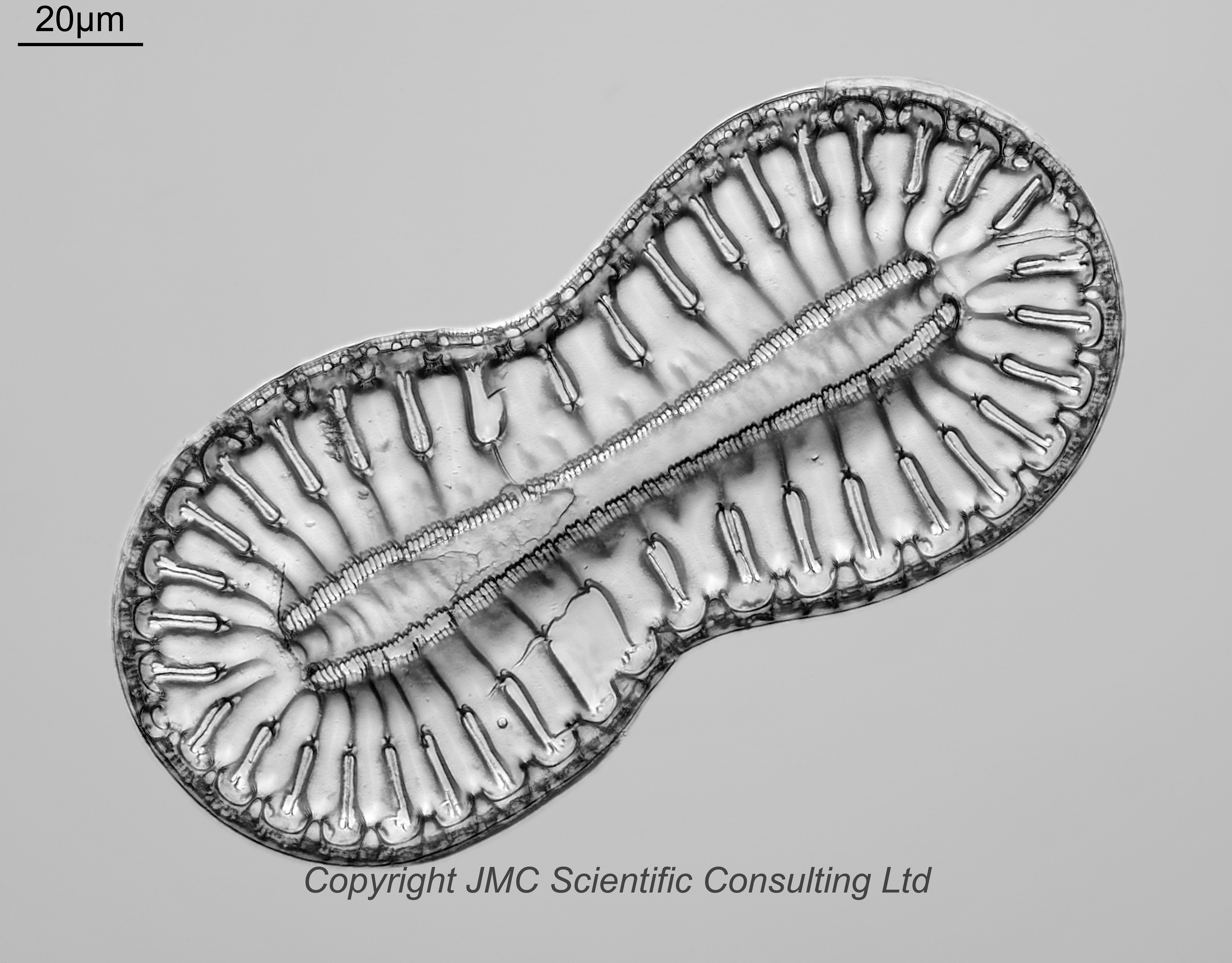
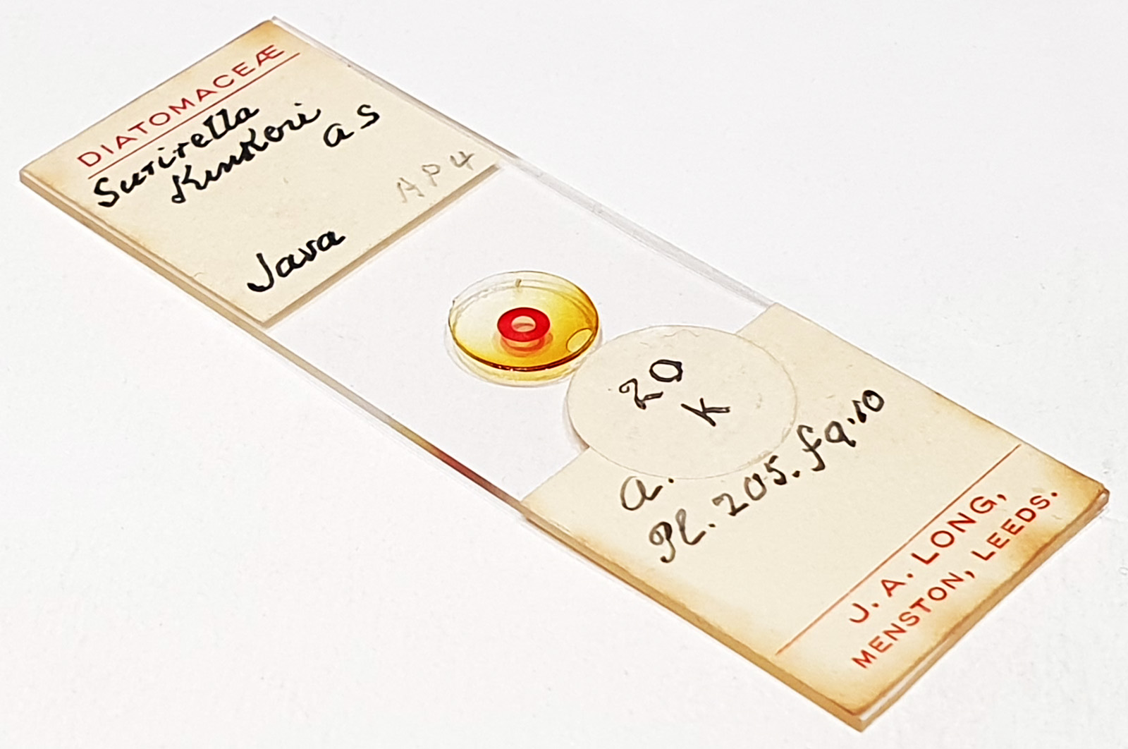
Surirella kinkeri from Java. Single example on the slide, along with a sponge spicule. Slide labelled as A. (presumably Schmidt) Plate 205, Figures 9,10 (see below for more on this). Prepared by JA Long. Olympus BHB microscope using 450nm LED light. 63x Leitz Pl Apo 1.4 objective, oil immersion. Olympus Aplanat Achromat condenser, oil immersion, oblique lighting. 2.5x Nikon CF PL photoeyepiece. Monochrome converted Nikon d850 camera. 51 images stacked in Zerene (Pmax). Not sure what the mountant is, but the diatom is higher than expected contrast (maybe it’s Hyrax?). However even for Hyrax it doesn’t look quite like I’d expect. Makes me wonder if the diatoms from some sites have different mineralogical makeup which then impacts their refractive index? However that is a question for another day. Whatever the reason, it was nice to image.
Surirella kinkeri A.W.F.Schmidt. Schmidt, A. (1897). Atlas der Diatomaceen-kunde. Leipzig. O.R. Reisland, Series V(Heft 52-53): pls. 205-212. Plate 205, Figures 9,10 (hence the writing on the label).