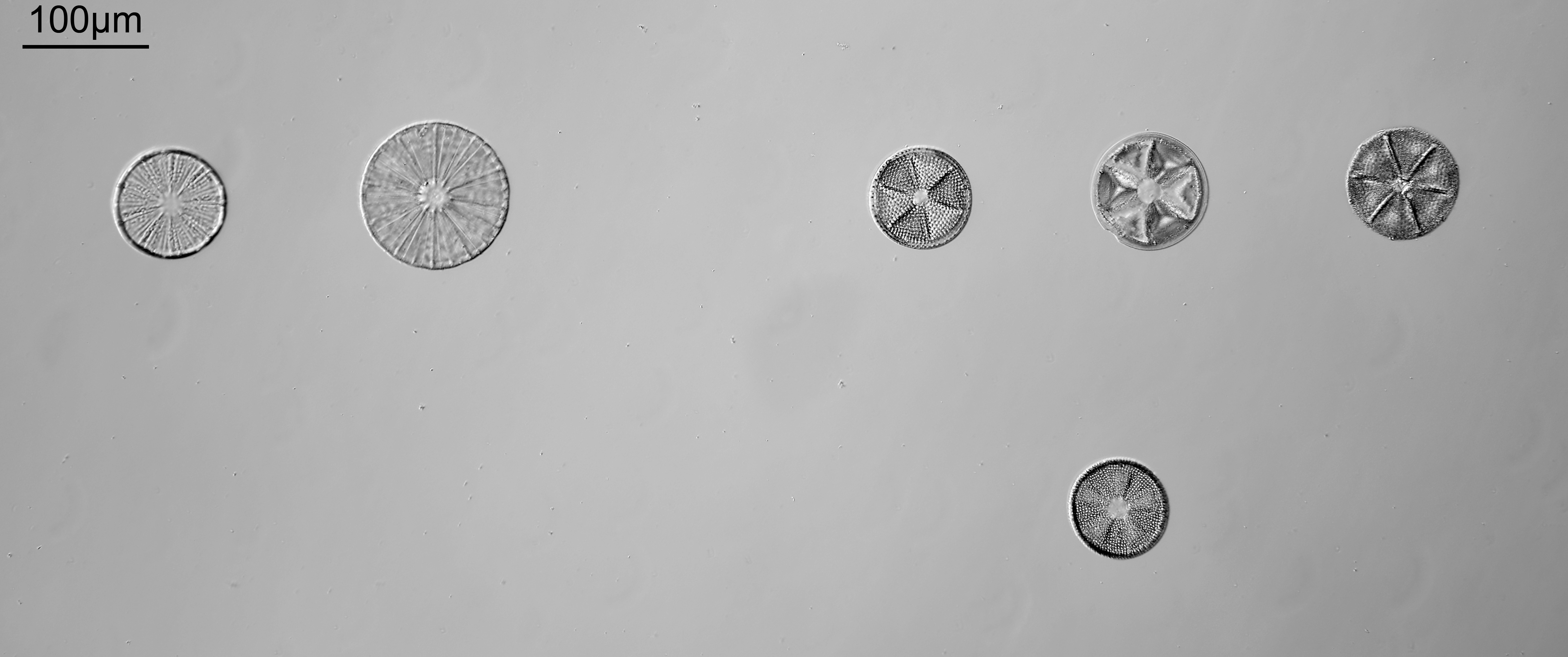
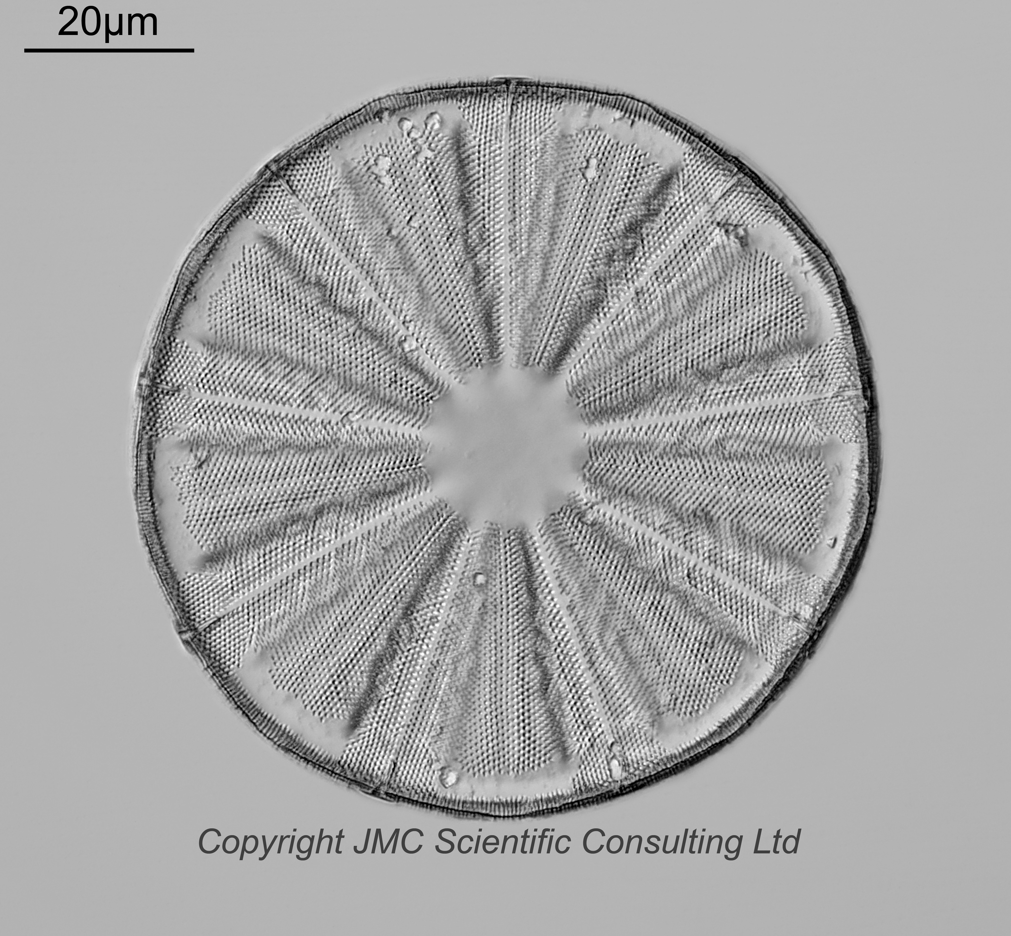
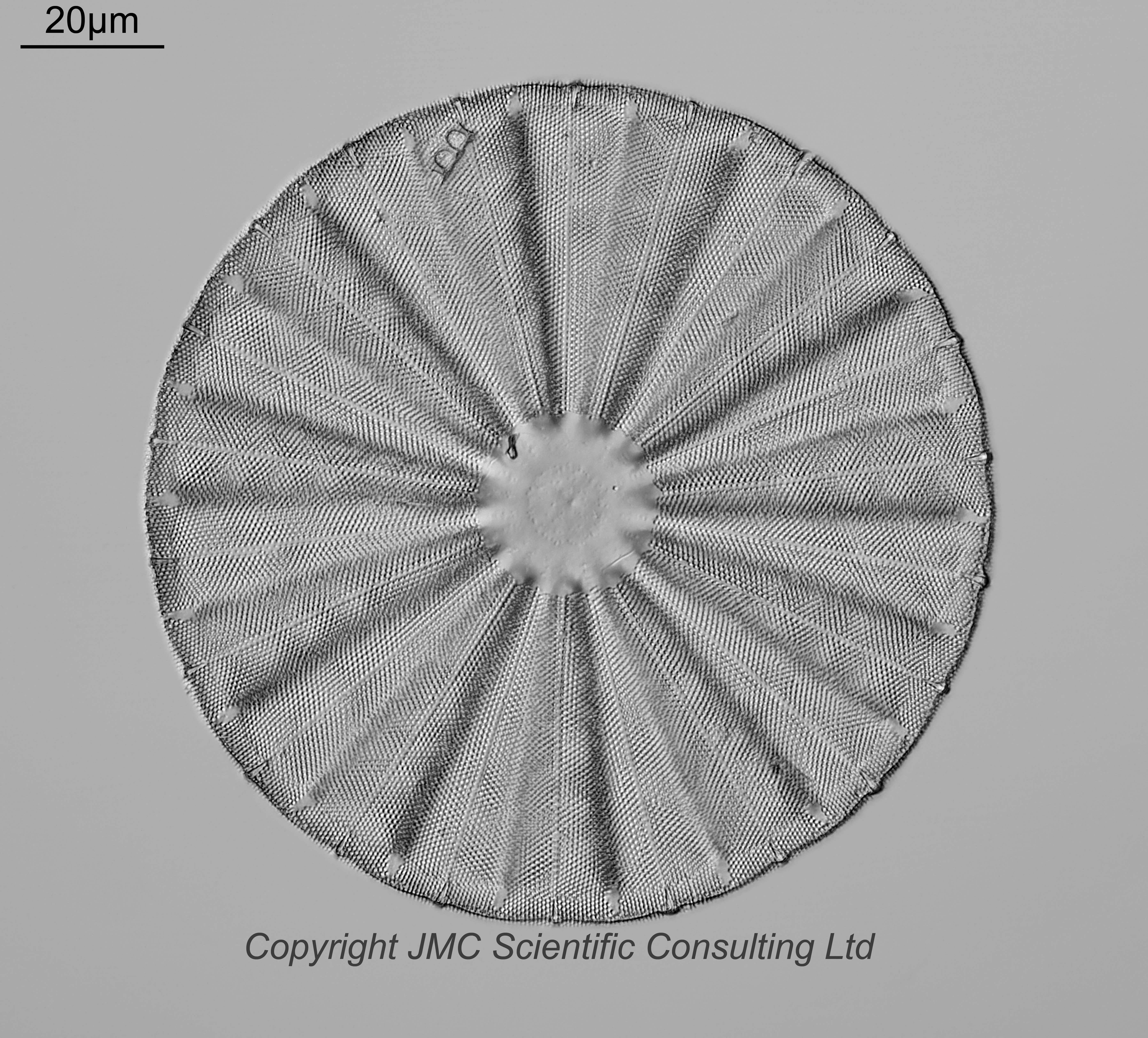
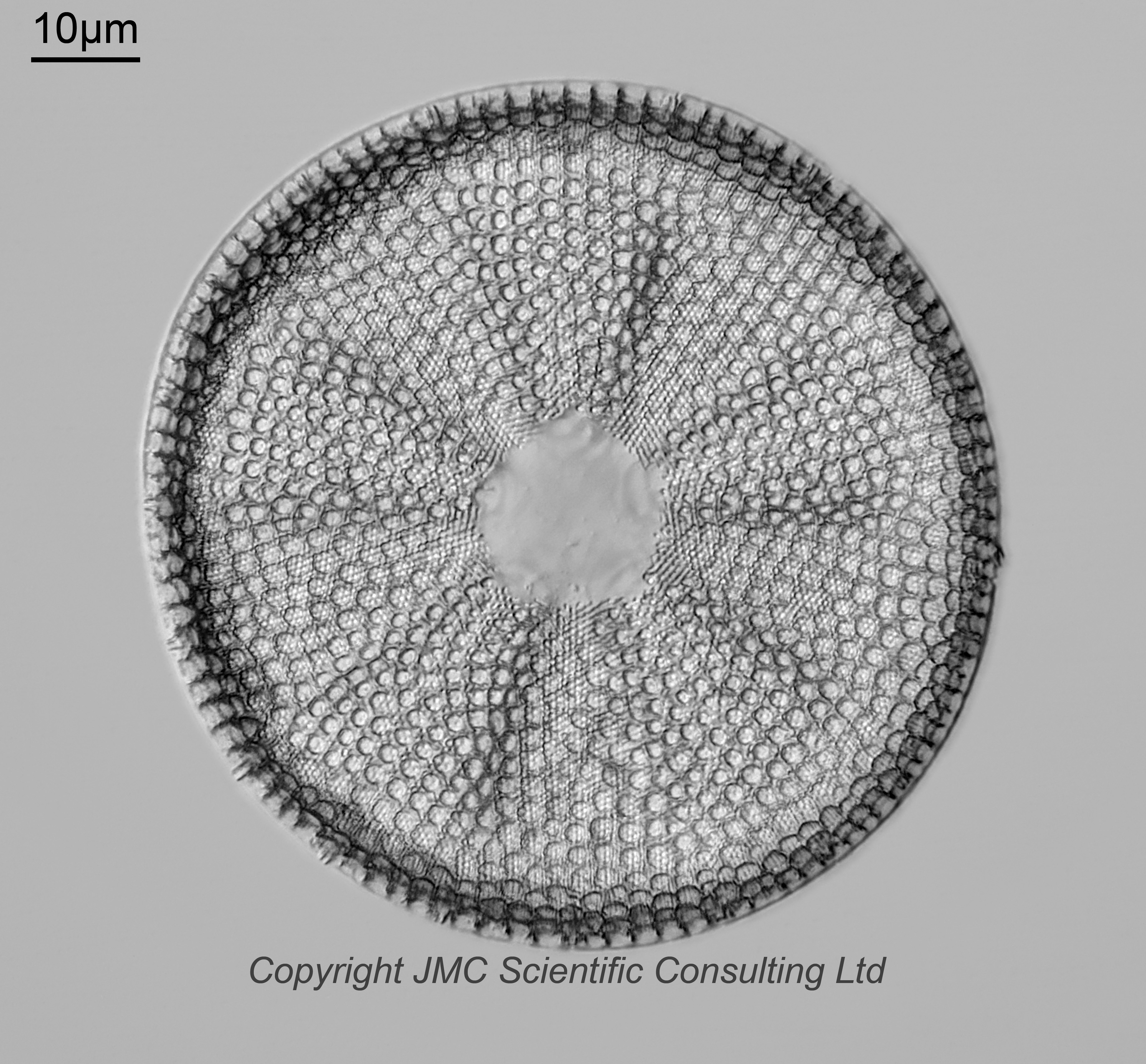
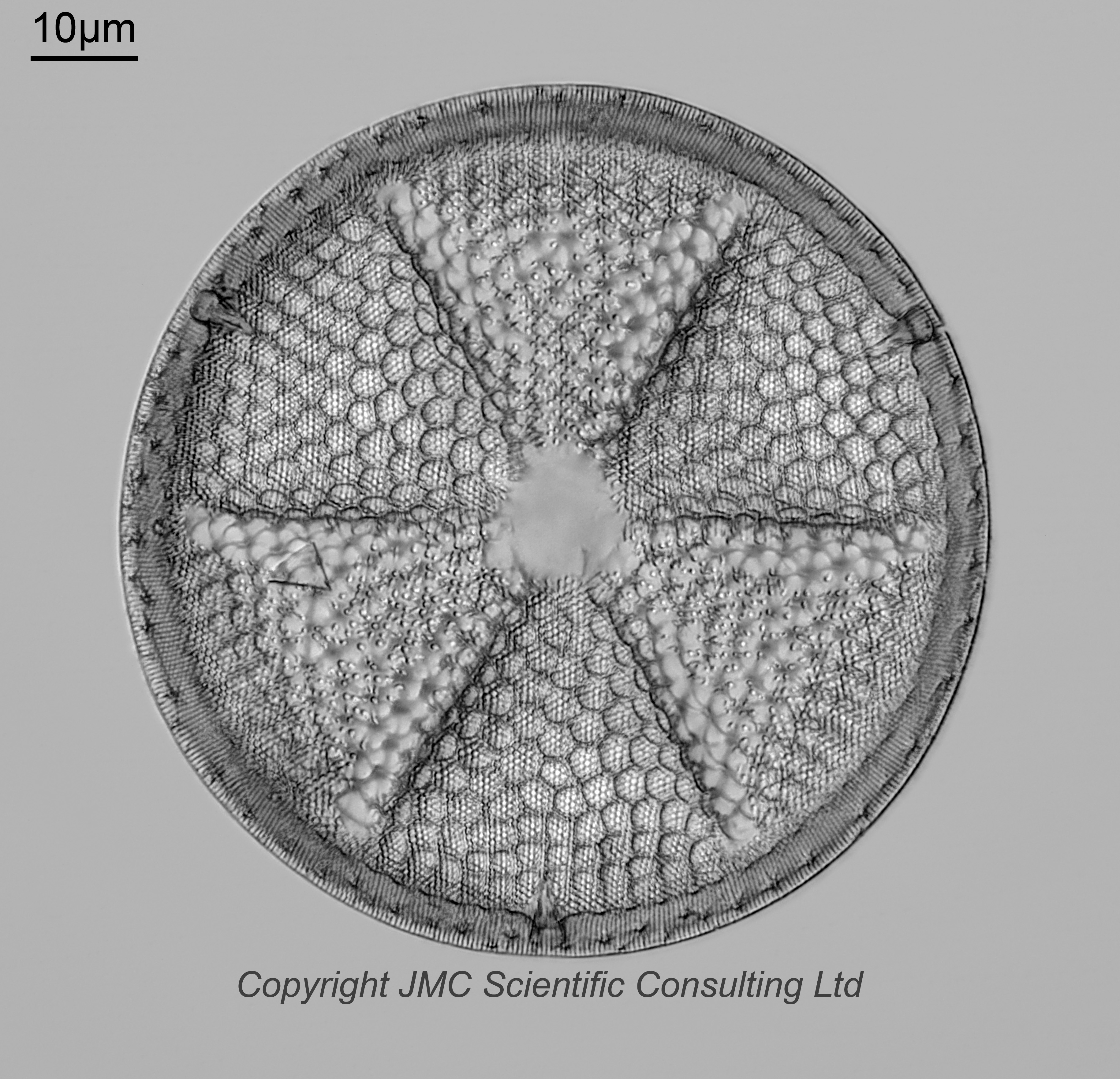
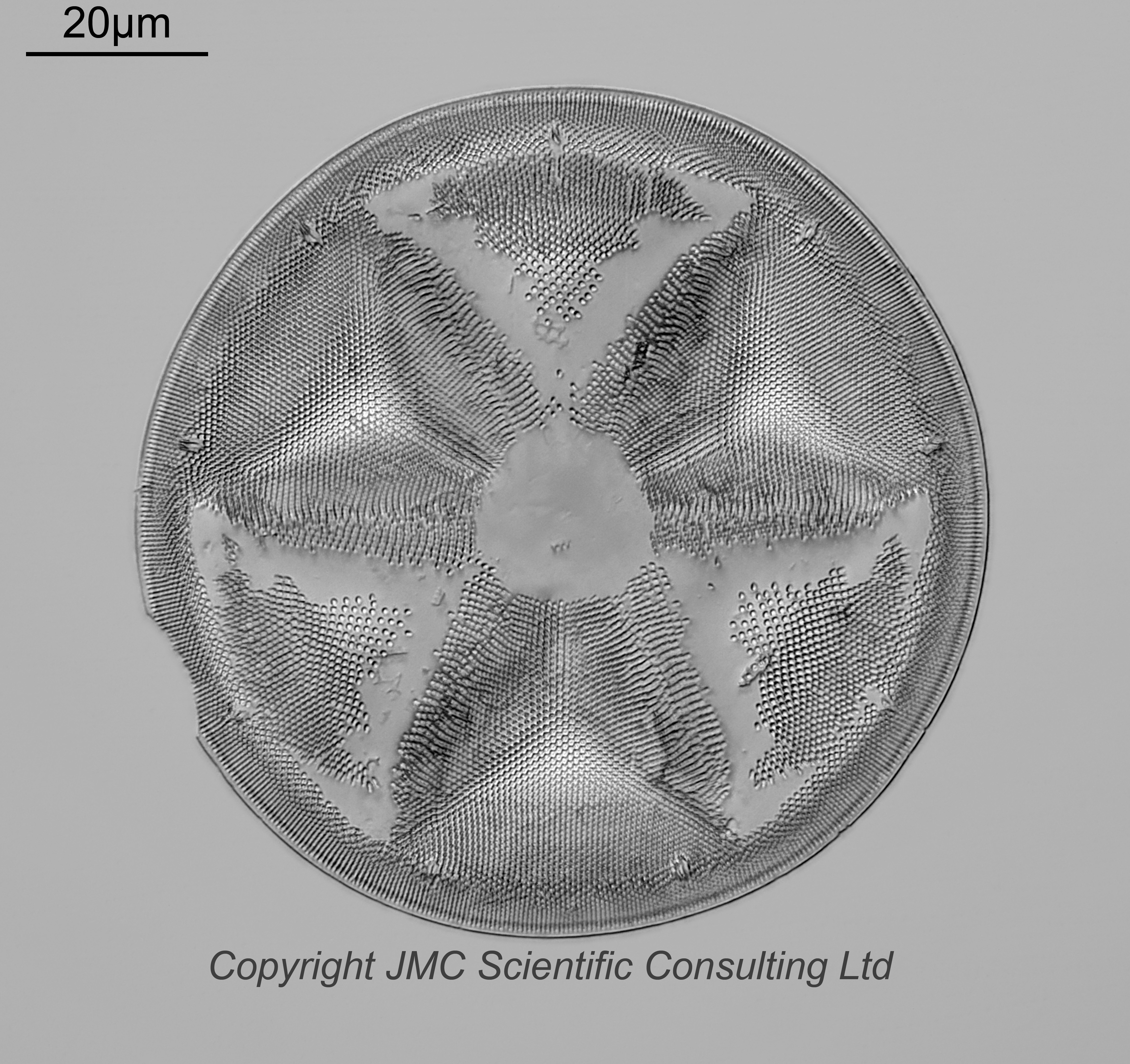
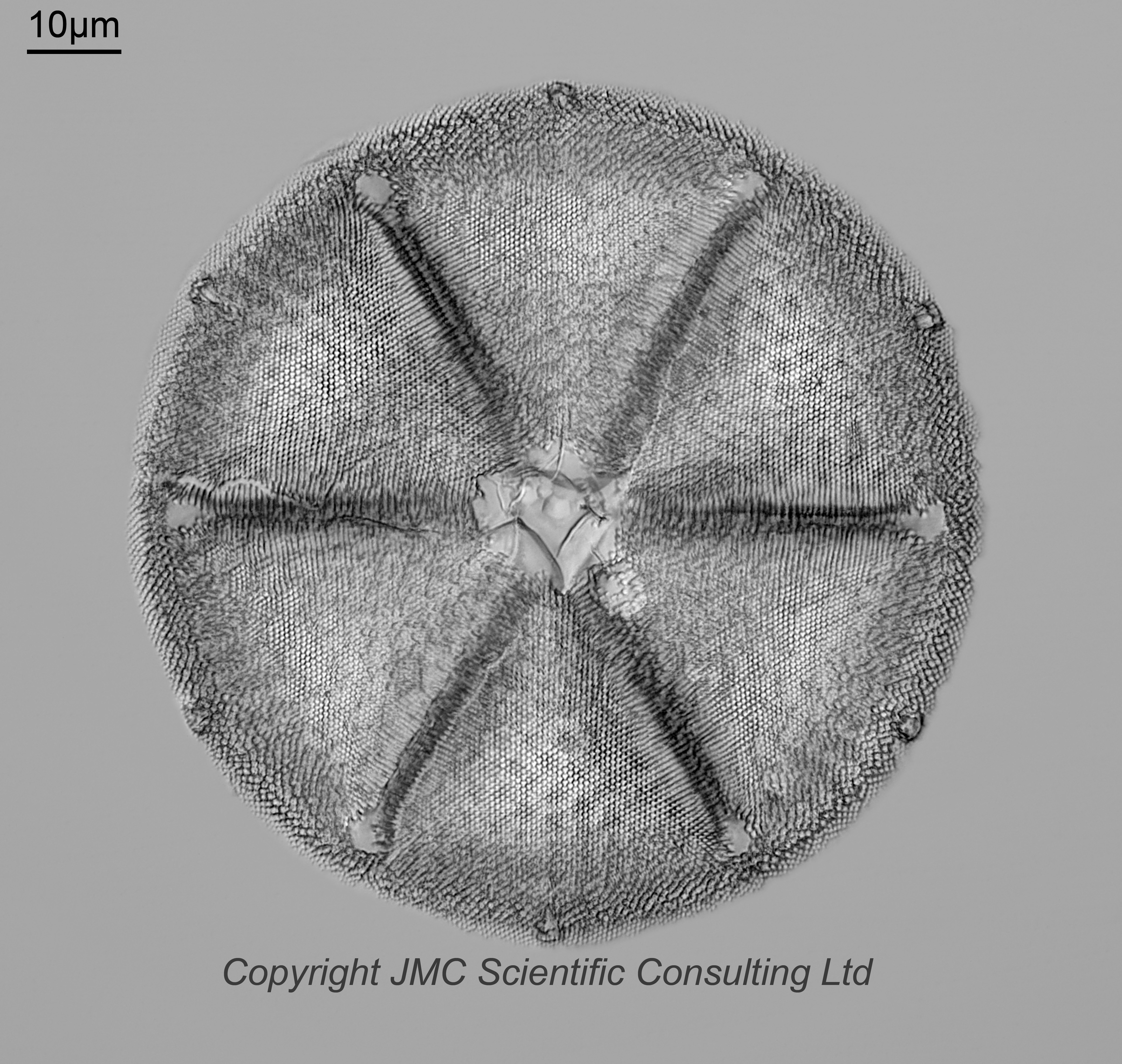
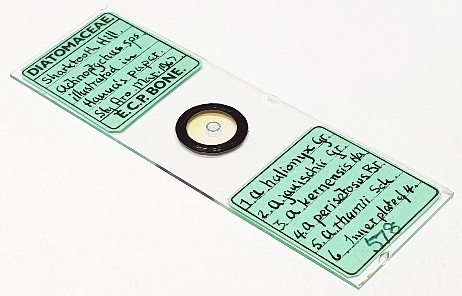
An arrangement of six Actinoptychus sps. from Sharktooth Hill, USA. Illustrated in Hanna’s paper (see later for details). One has moved from its original position. Sty.Aro. (Styrax Aroclor) mountant, and dated March 1967. Prepared by ECP Bone. Olympus BHB microscope using 450nm LED light. Imaged inidividually using a 63x Leitz Pl Apo 1.4 objective, oil immersion. Olympus Aplanat Achromat condenser, oil immersion, oblique lighting. 2.5x Nikon CF PL photoeyepiece. Monochrome converted Nikon d850 camera. Images stacked in Zerene (Pmax).
The paper referred to above is; The diatoms of Sharktooth Hill, Kern County, California, by G Dallas Hanna, published in Proceedings of the California Academy of Sciences, in 1932, in volume 20, pages 161-263.
Left to right in the arrangement;
1. Actinoptychus halionyx. 15 images stacked. Plate 2, Figure 4 in Hanna’s Sharktooth Hill paper.
2. Actinoptychus janischii. 19 images stacked. Plate 3, Figure 1 in Hanna’s Sharktooth Hill paper.
3. Actinoptychus kernensis. 27 images stacked. Plate 3, Figures 2 and 3 in Hanna’s Sharktooth Hill paper. This one has moved from its original position in the arrangement.
4. Actinoptychus perisetosus. 50 images stacked. Plate 4, Figures 1 and 2 in Hanna’s Sharktooth Hill paper.
5. Actinoptychus thumii. 49 images stacked. Plate 4, Figures 3 and 4 in Hanna’s Sharktooth Hill paper.
6. Actinoptychus perisetosus (inner plate). 43 images stacked. Plate 4, Figures 1 and 2 in Hanna’s Sharktooth Hill paper.
Interestingly, they are arranged in the same order they appear in the article – alphabetically – apart from the inner plate of A. perisetosus which has been placed at the right hand side of the arrangement.