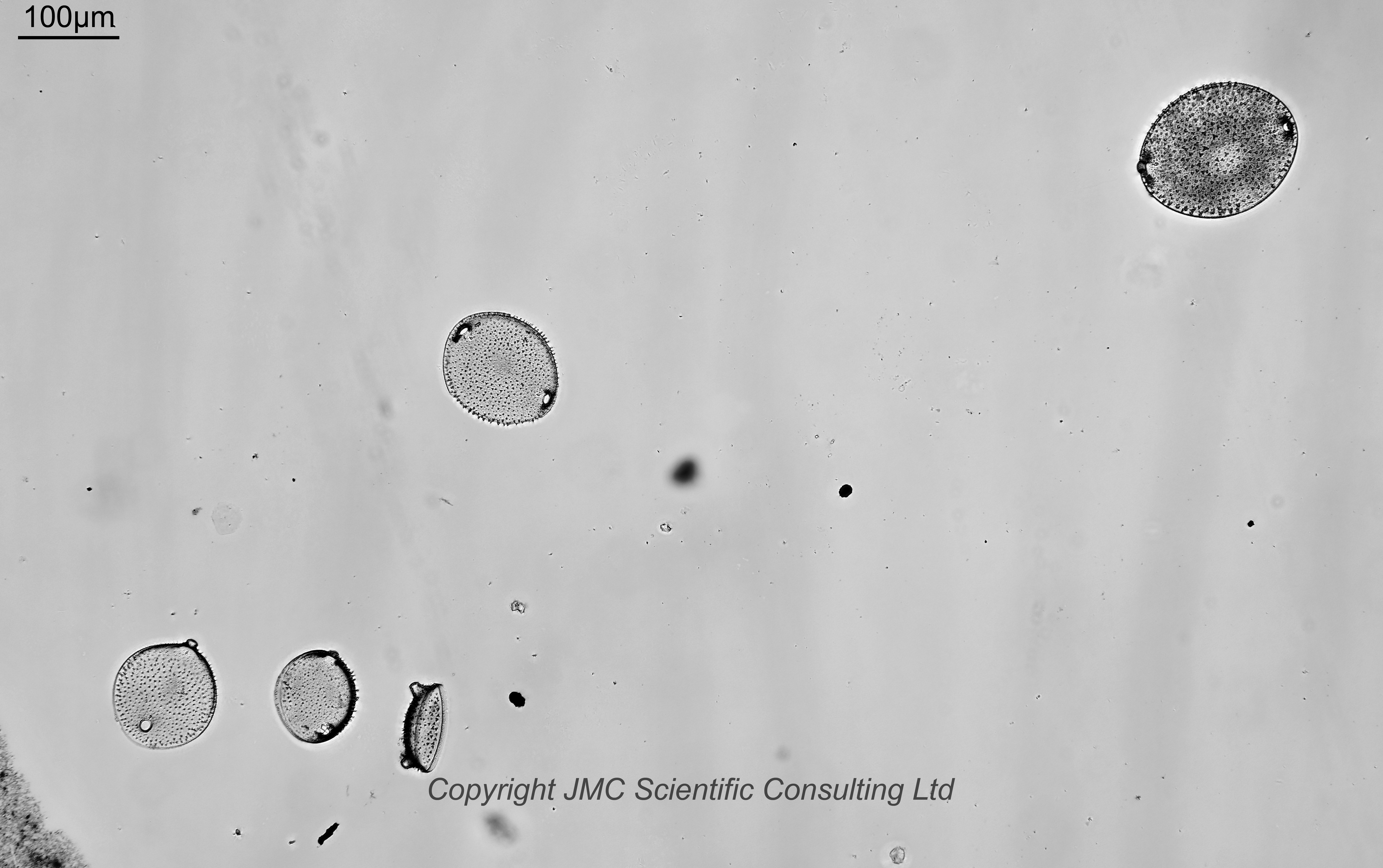
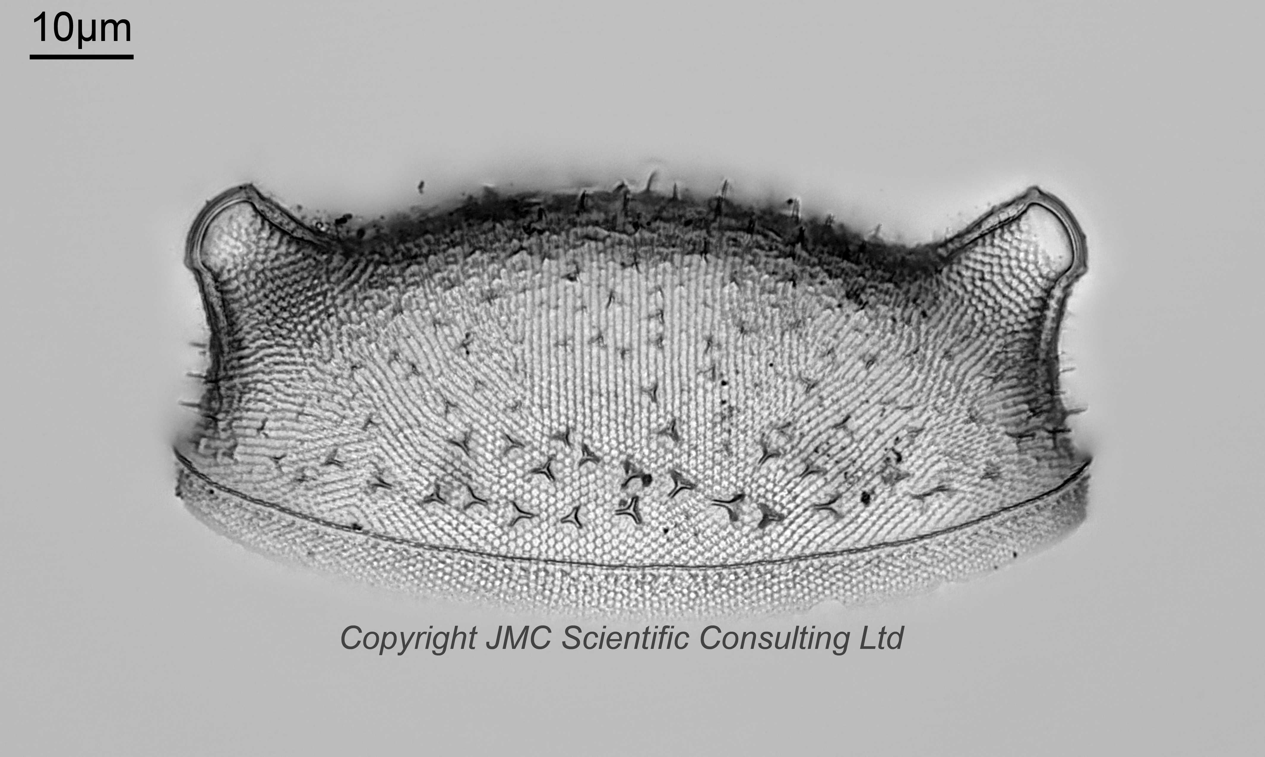
A slide of Biddulphia echinata from Fiji. Different orientations. Some have moved away from the middle of the coverslip. Prepared by Arthur Cottam. Olympus BHB microscope using 450nm brightfield LED light. Wider arrangement imaged using a 10x Nikon Plan Apo NA 0.45 objective. Individual diatoms imaged using a 63x Leitz Pl Apo NA 1.40 oil immersion objective. Olympus Aplanat Achromat condenser, oil immersion, brightfield lighting. 2.5x Nikon CF PL photoeyepiece. Monochrome converted Nikon d850 camera.
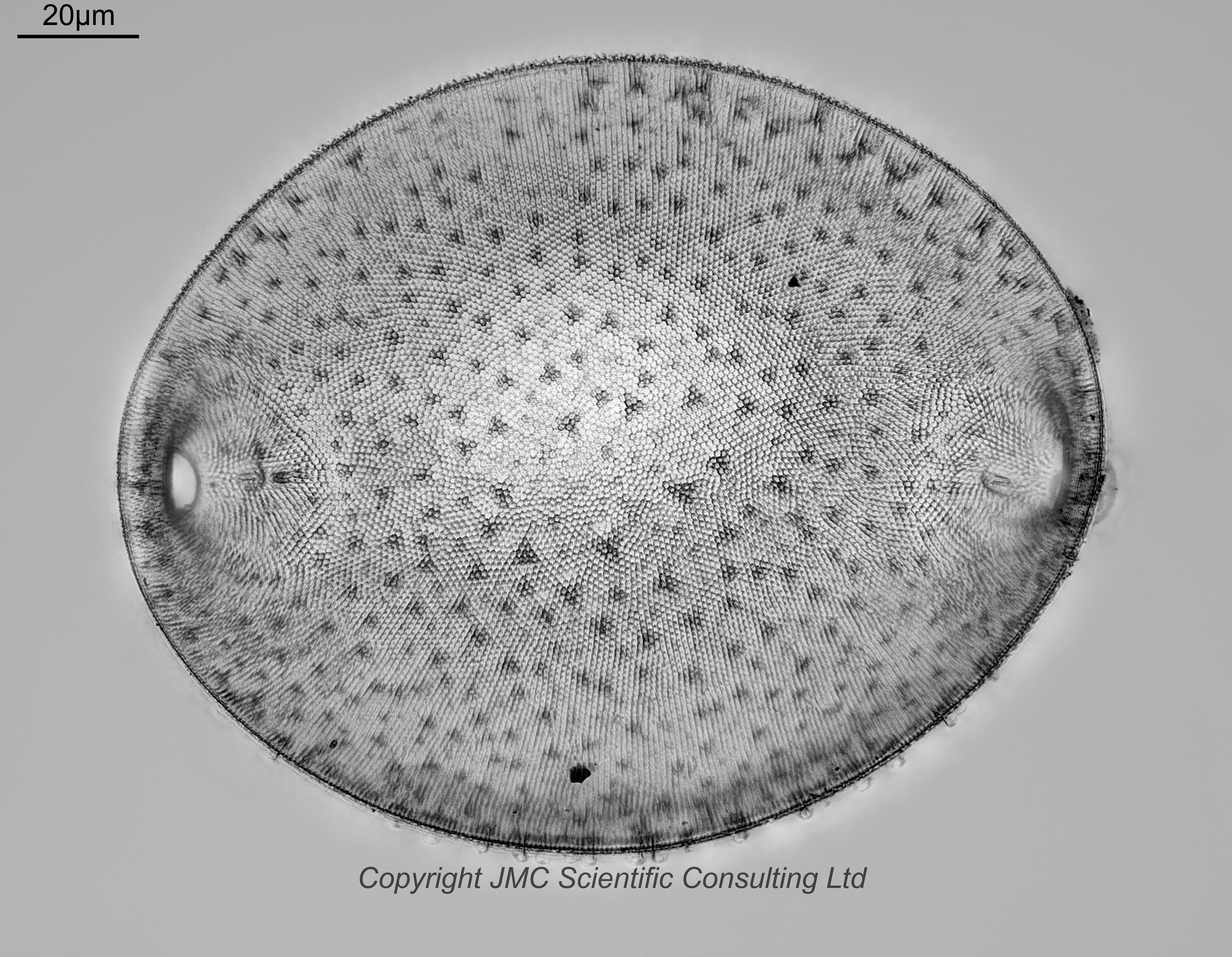
Two examples are shown at higher resolution. One has the diatom flat to the coverslip, but with the rim facing upwards towards the camera (it is showing the inside/underside of the diatom). It is a stacked image, one of the largest stacks I have done due to the very 3D nature of the diatom, 130 images in Zerene (Pmax). Some of the subtle details visible in the individual frames have been lost during stacking, but these are more easily seen in the crops from single frames.
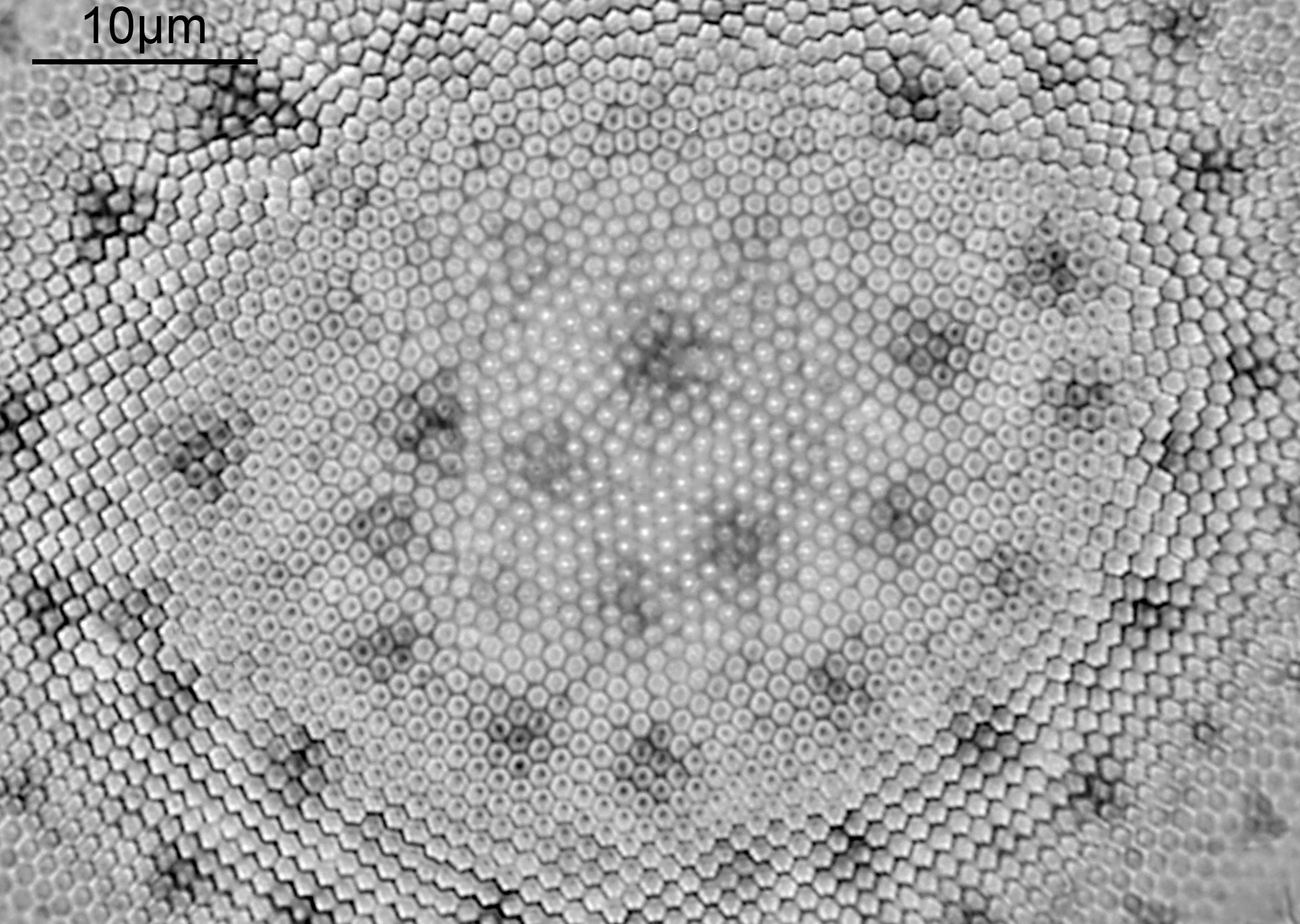
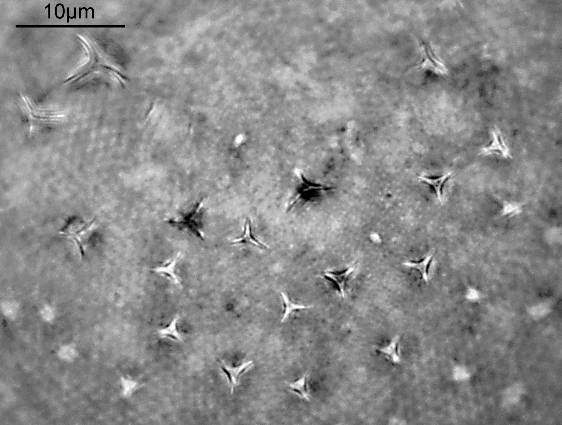
The second one shows the outside of the diatom view from the side (and looks like the ‘head’ of a Dalek to me). The three pointed star cross section of the spines is nicely visible now as is the really intricate surface structure. This was a stack of 100 images in Zerene (Pmax). Overall a very pretty diatom, but a challenge to image.
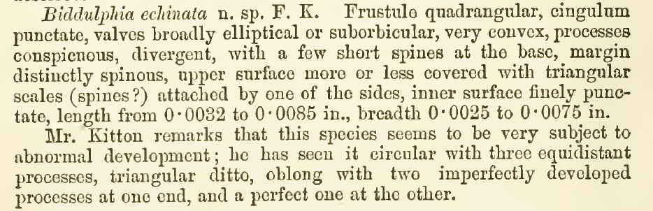
It is written about in Kitton, F. (1888). New species of Biddulphia from Fiji. Journal of the Royal Microscopical Society, 1888: p. 466, and I have included a screen grab of that here.