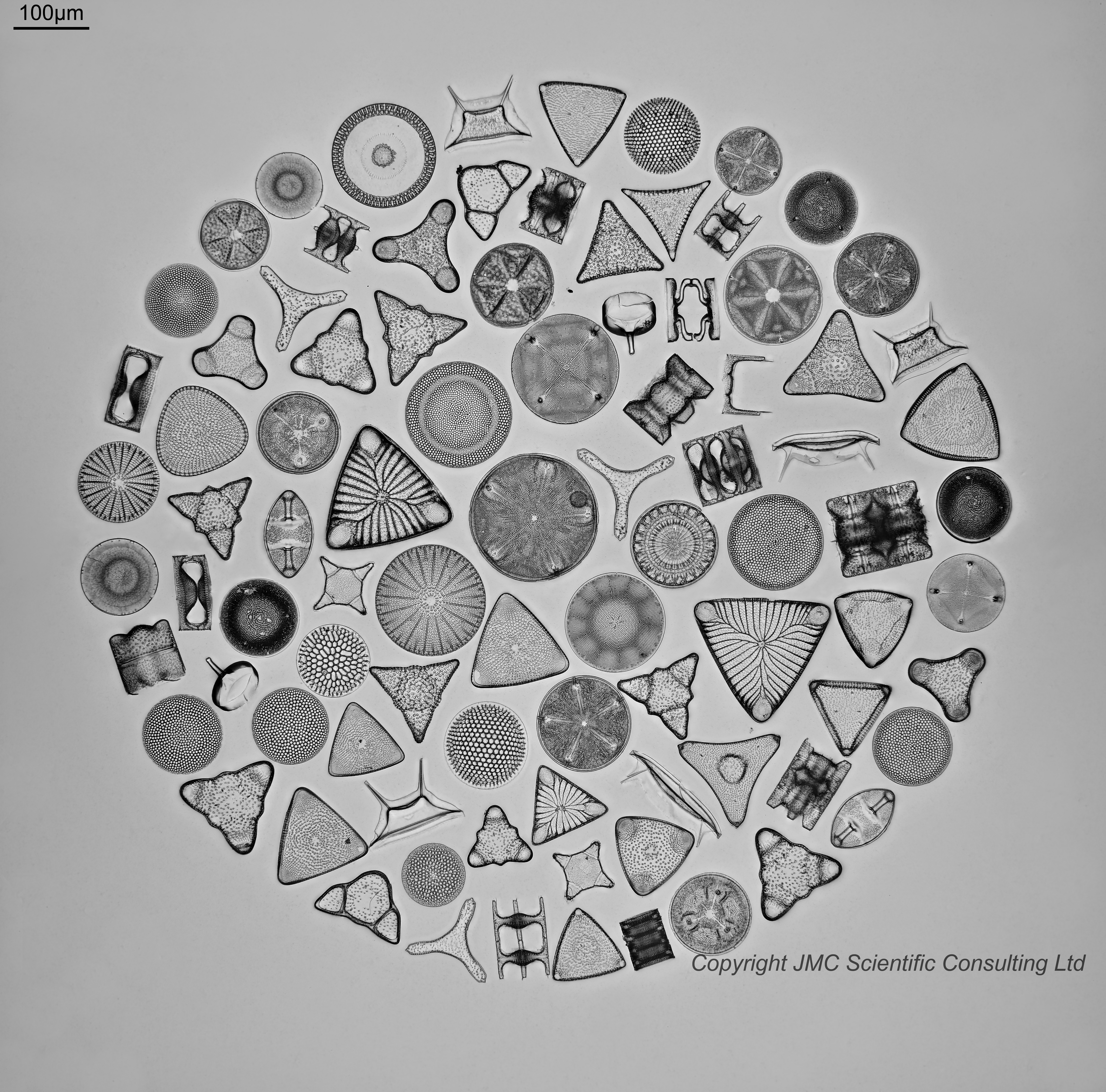
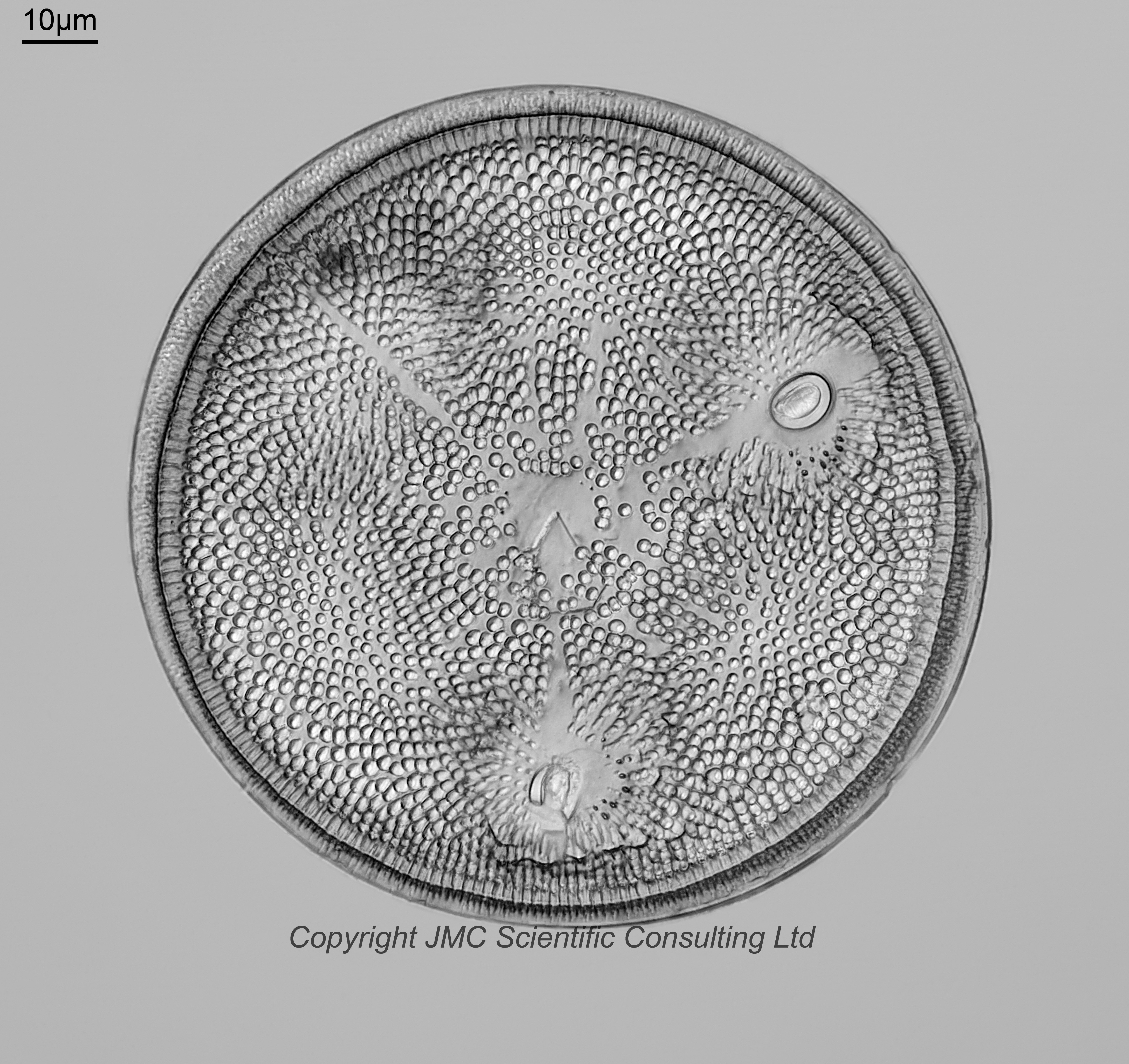
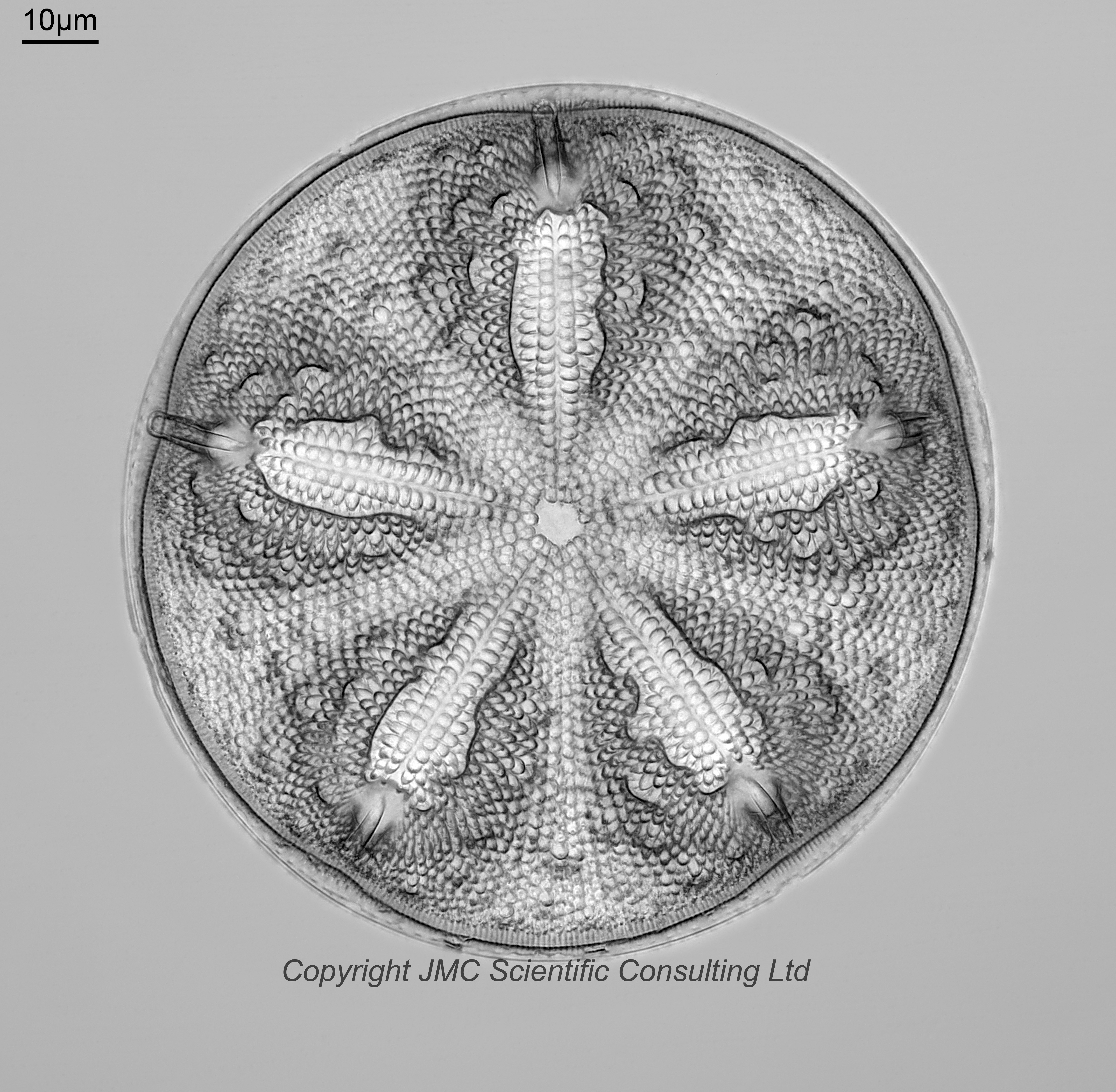
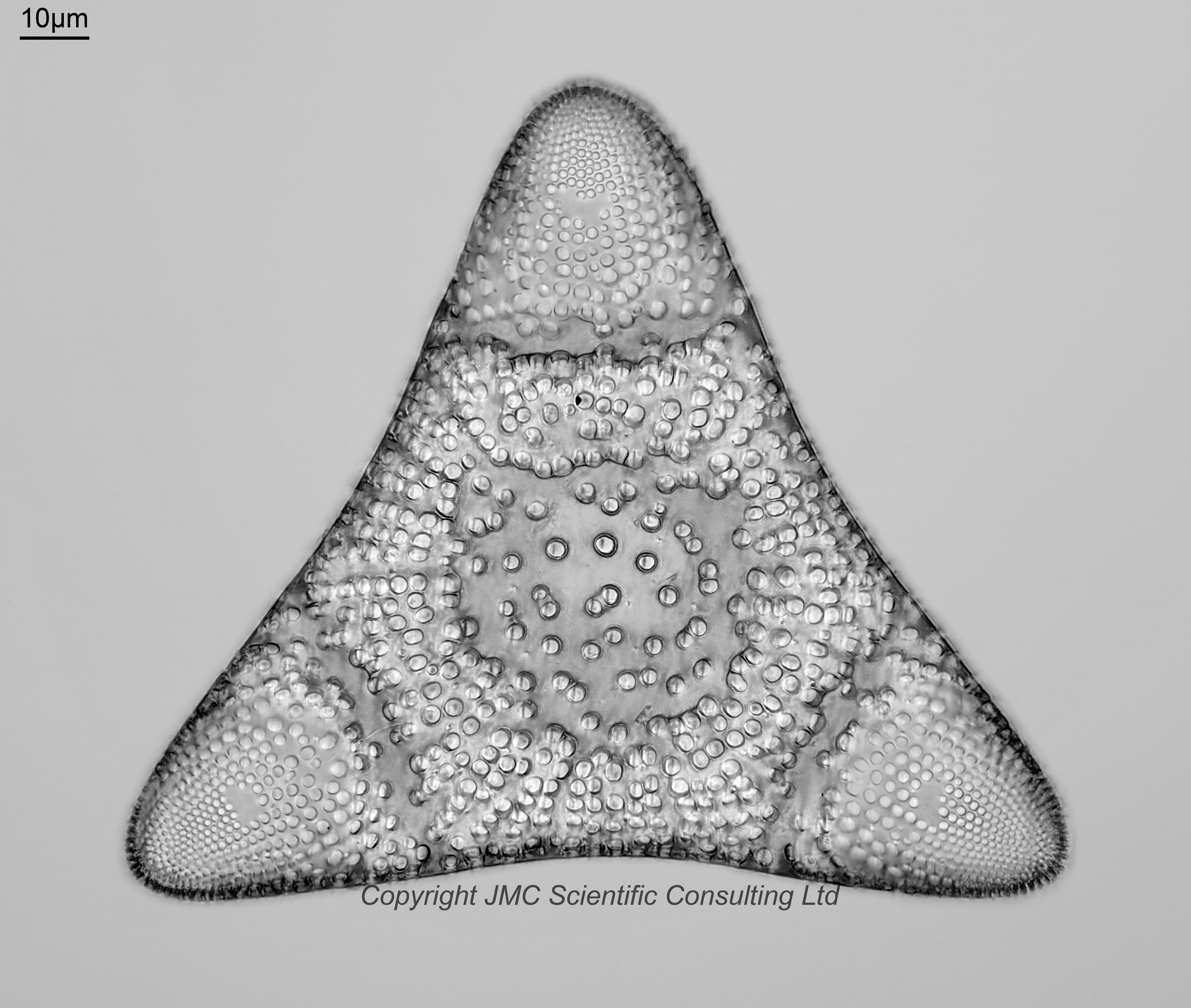
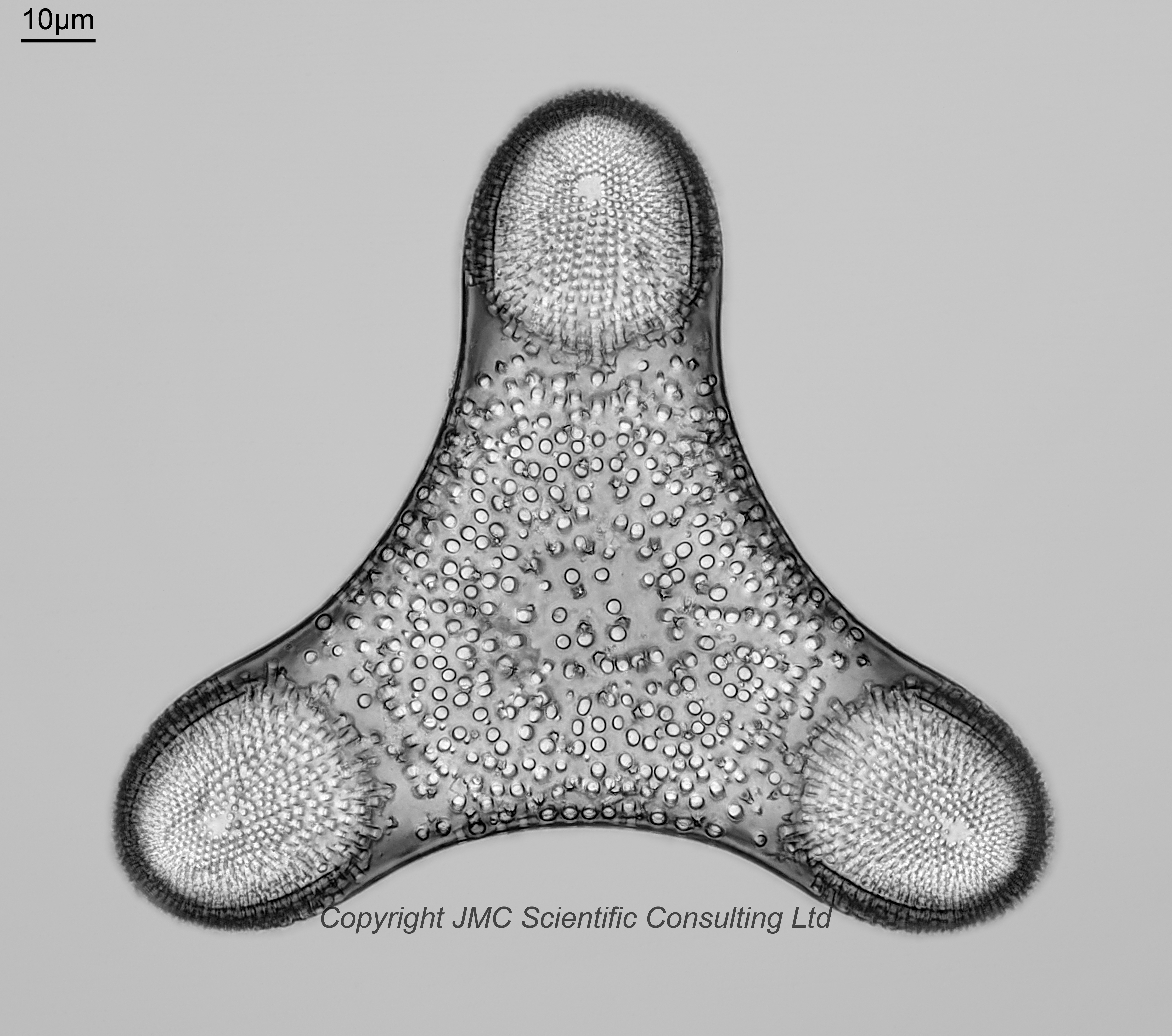
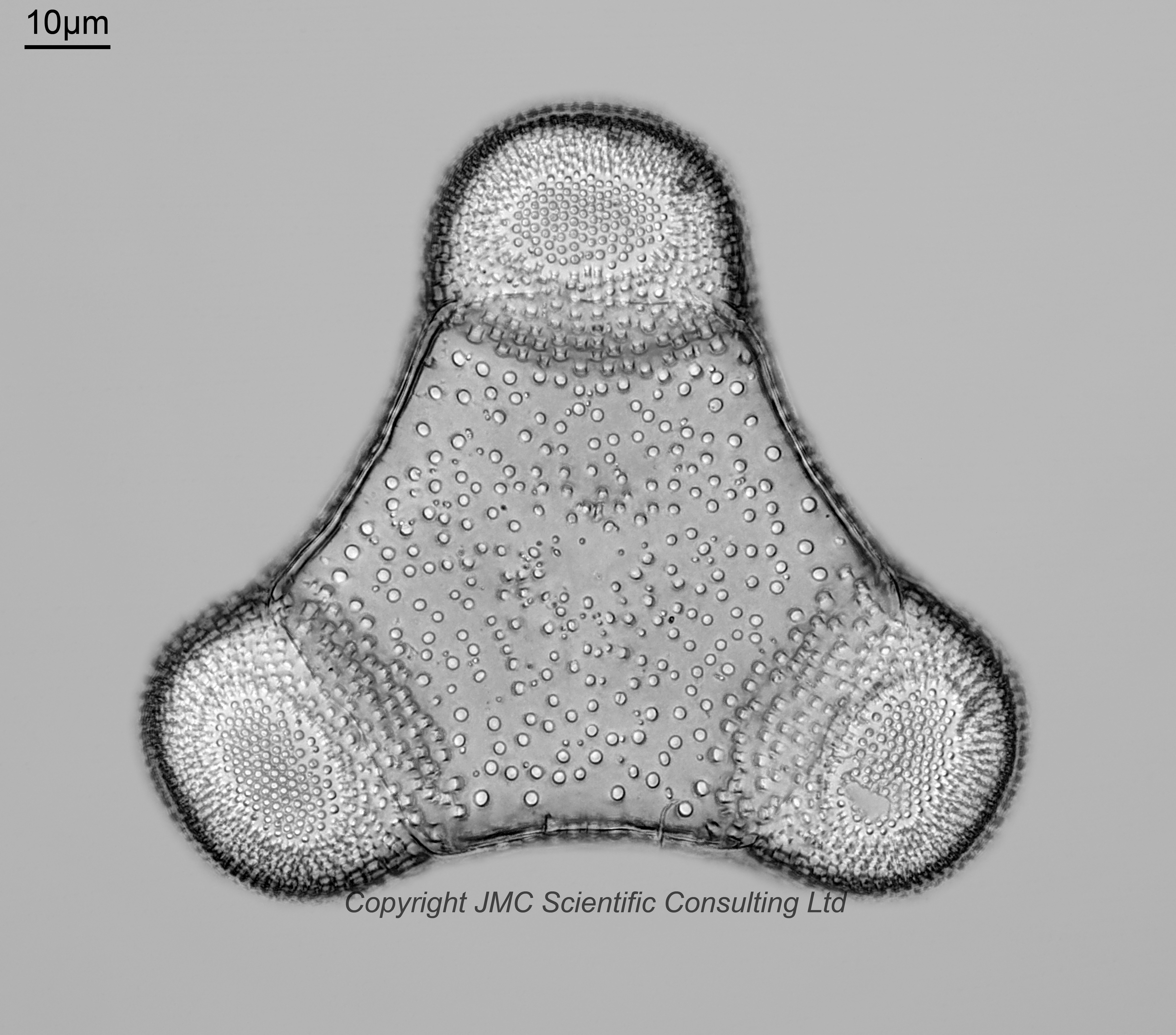
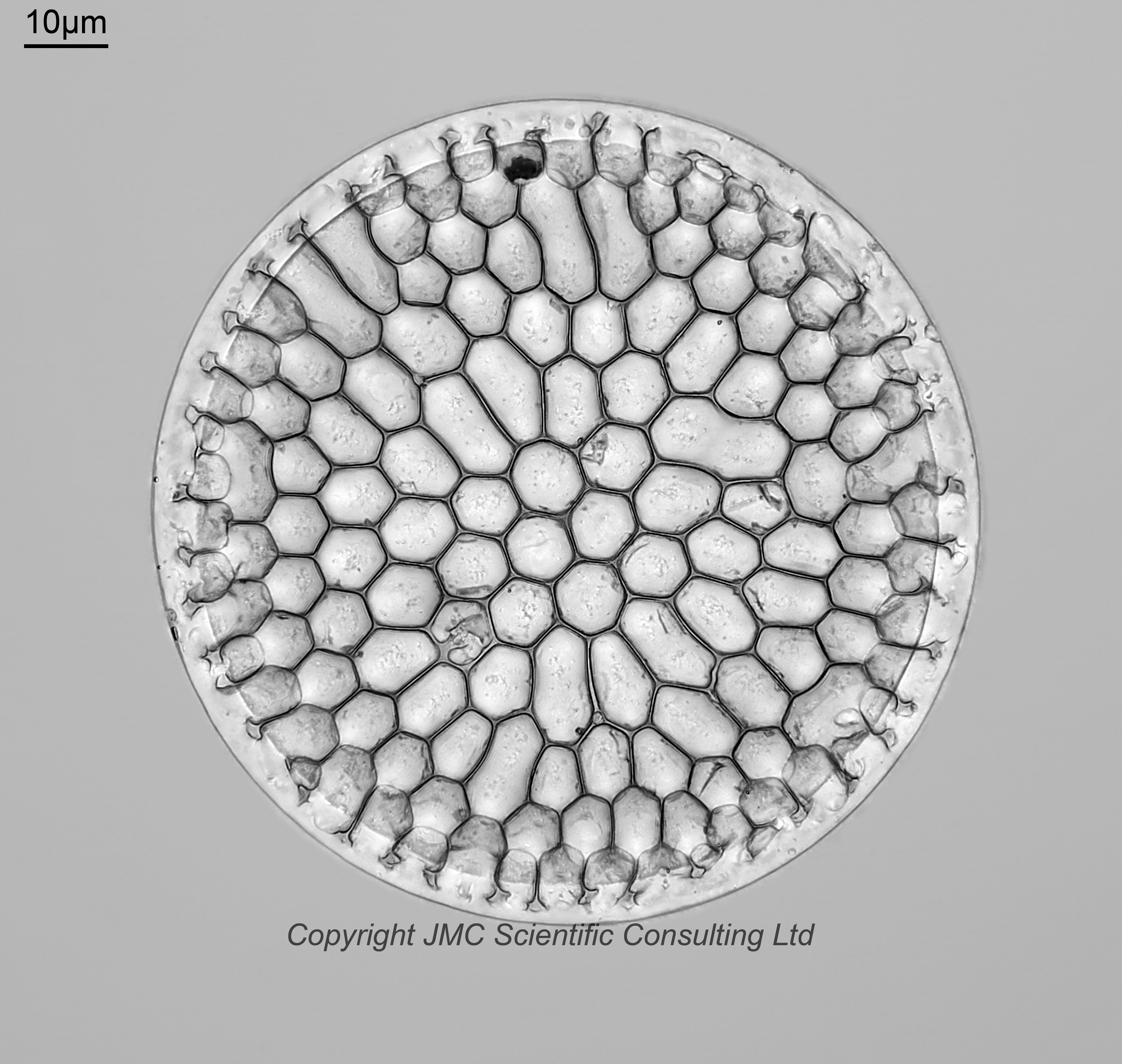
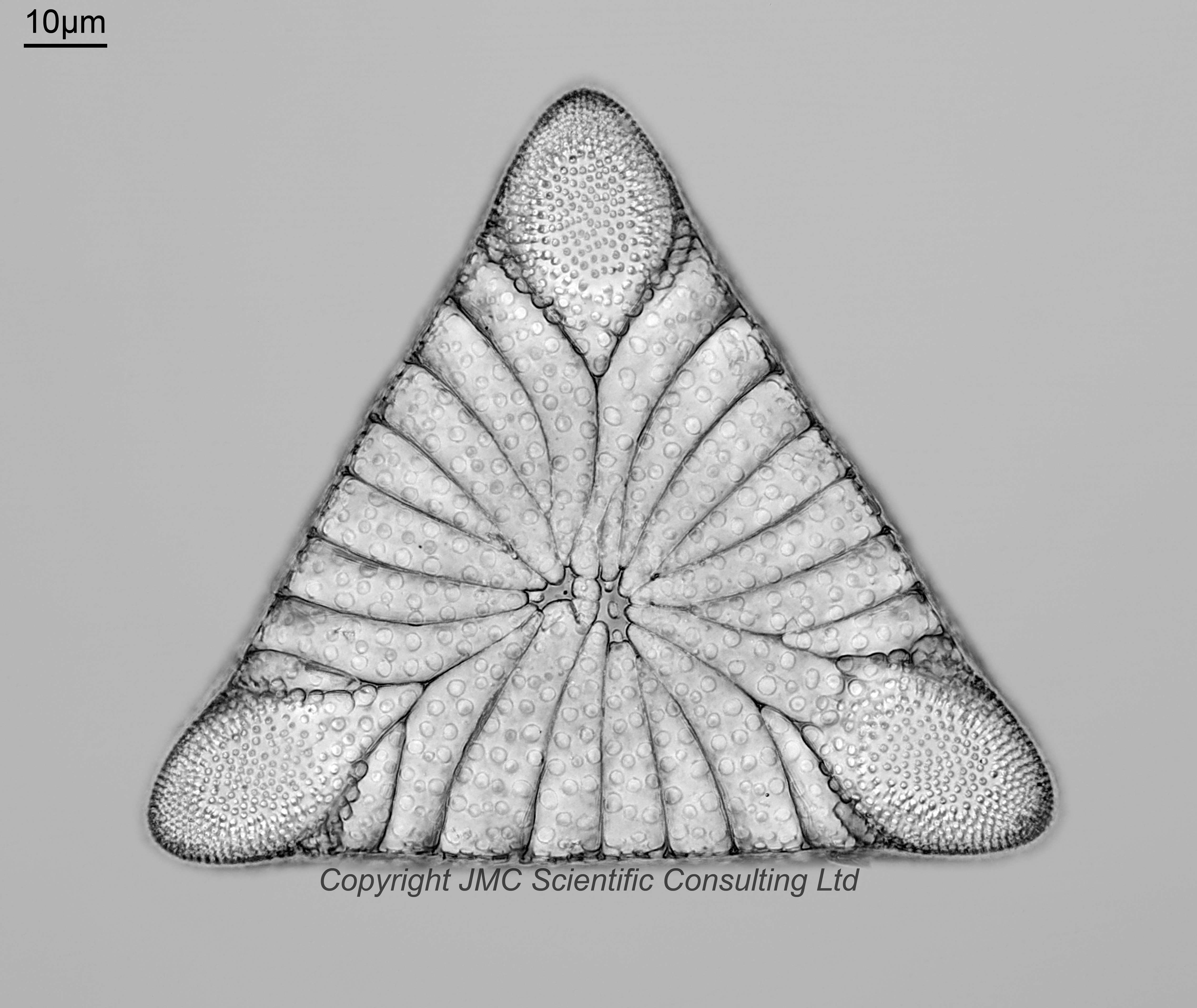
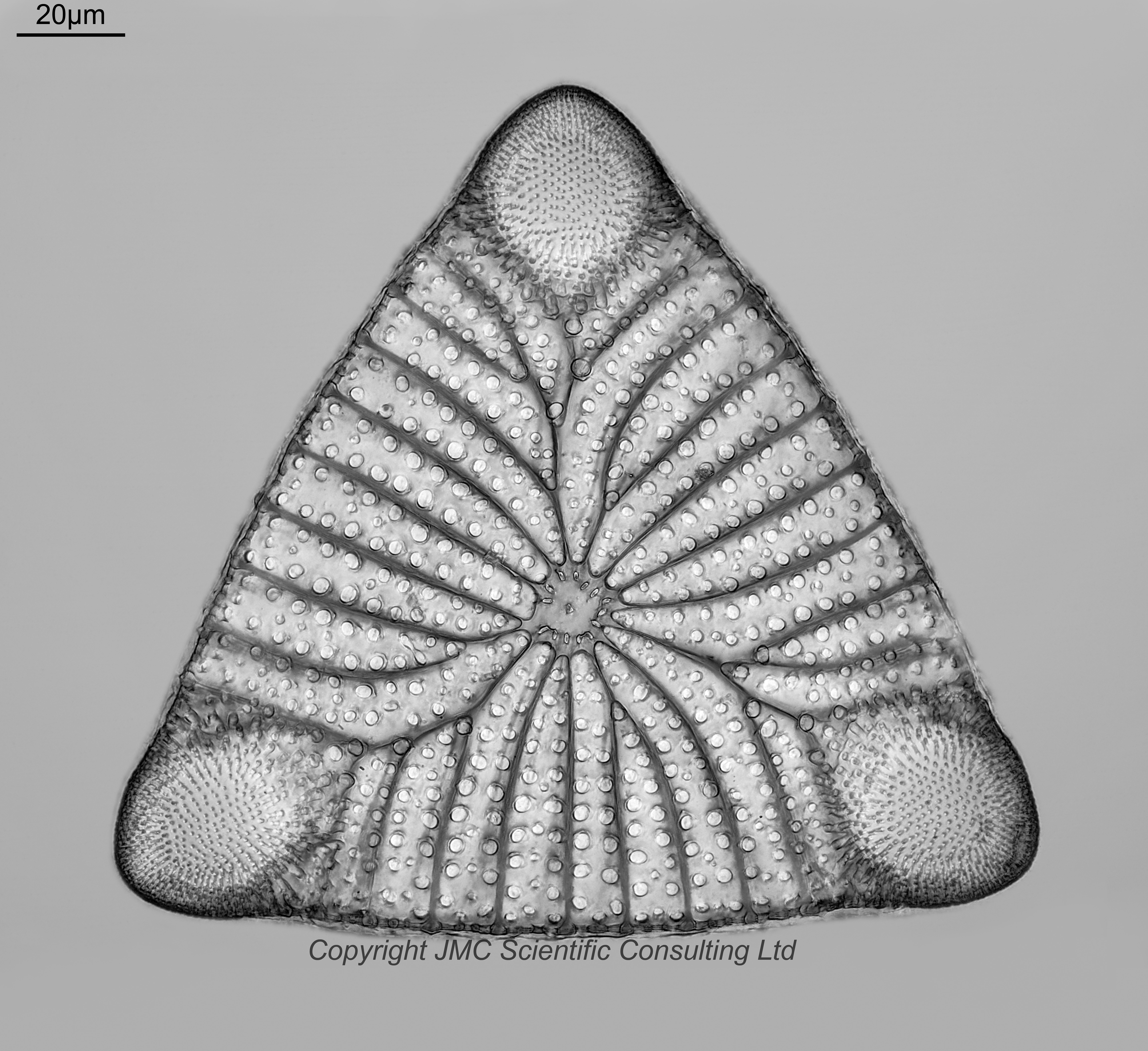
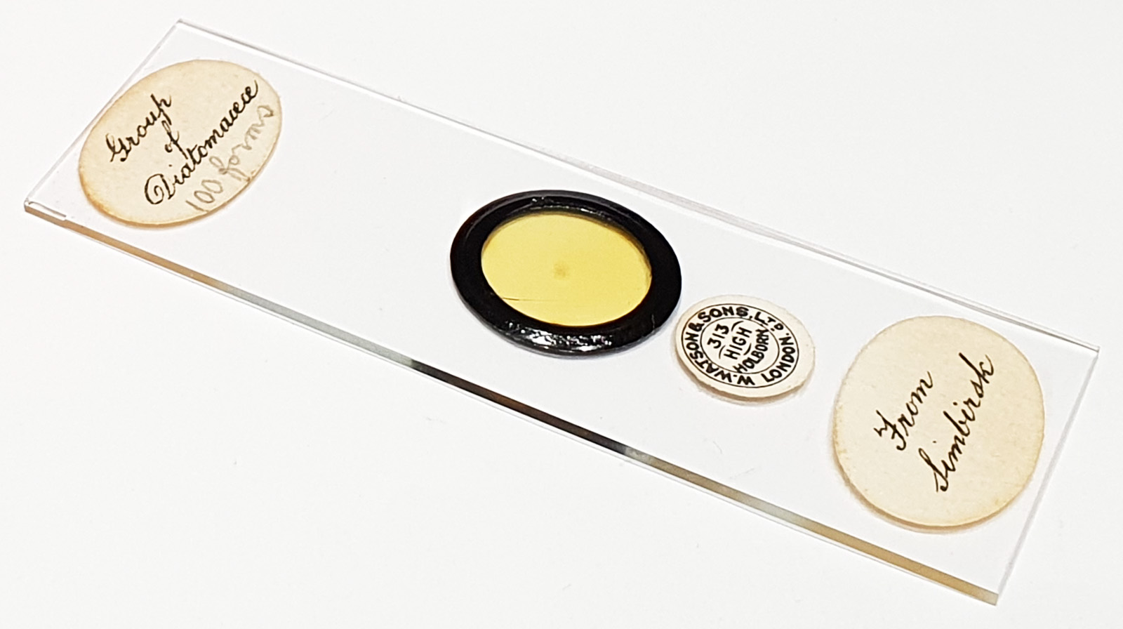
A really nice 100 form arrangement from Simbirsk, Russia, with some very well preserved diatoms. Prepared by Watson and Sons Ltd. Olympus BHB microscope using 450nm LED light. 63x Leitz Pl Apo 1.4 objective, oil immersion. Olympus Aplanat Achromat condenser, oil immersion, brightfield or oblique lighting. 2.5x Nikon CF PL photoeyepiece. Monochrome converted Nikon d850 camera. Image stacks prepared in Zerene (Pmax).
Some of my identifications are very tentative here, so please keep that in mind when reading the rest of this post.
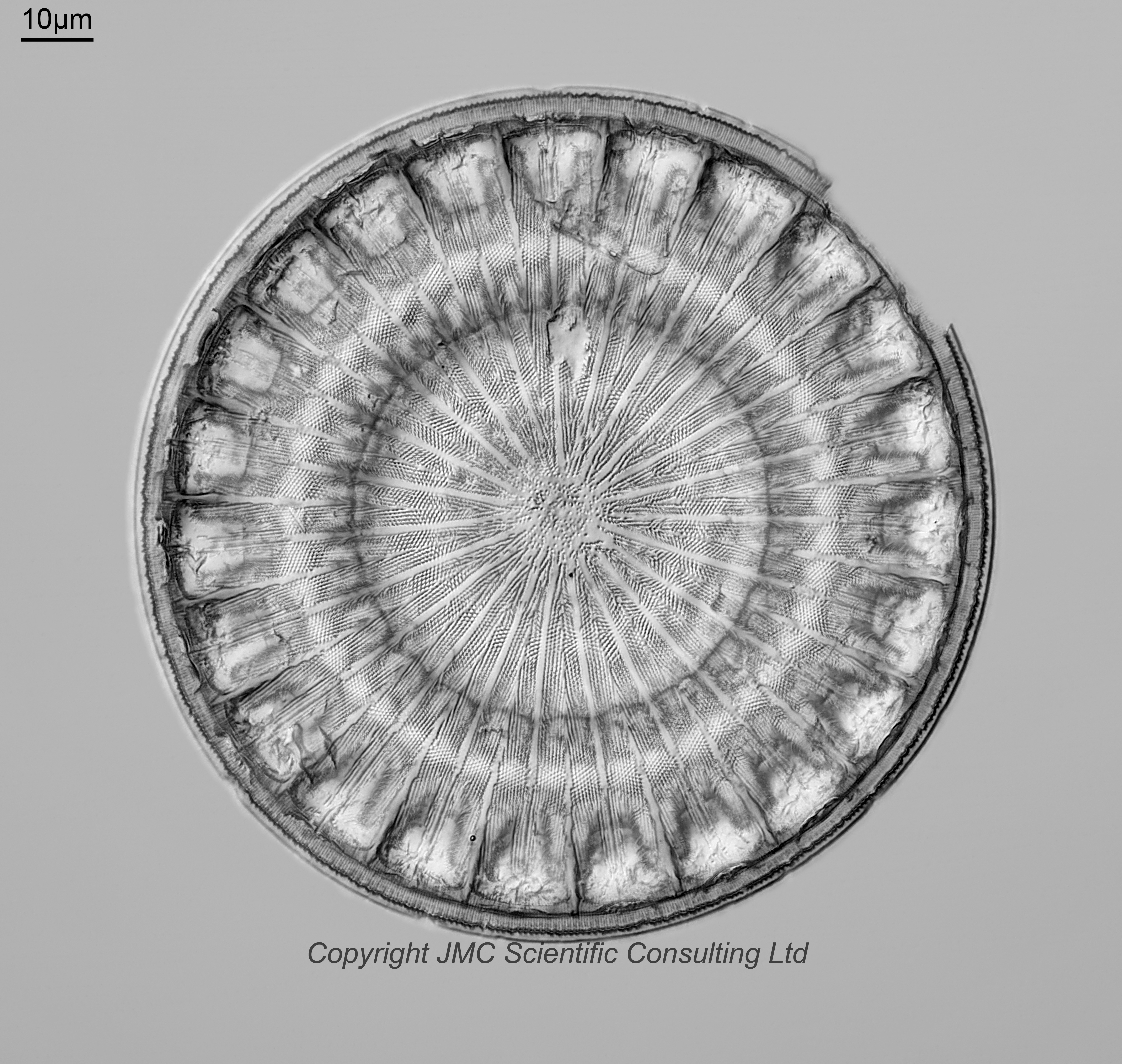
Lepidodiscus sp. This could be Lepidodiscus elegans or Lepidodiscus sublimis. Oblique lighting. 53 images in Pmax. Some of the dots have ‘blended’ together in the stacked image just outside the middle region, and on the bit just inside the rim. Dots are visible in the original images in more places than the stack. There is an interesting bit by Barber (Some Further Works by Horace George Barber, available here) about the trouble with identifying examples of this genus; “The identification of the species of the genus Lepidodiscus is most difficult due to the very poor illustrations to hand for this purpose. i.e O. Witt. Simbirsk Diatoms Pl.VII Fig. 6., Lefebure. Atlas of Diatoms Pl.8. Fig.5., H. Coupin. Atlas of Diatoms Pl.294. Fig.J. These three authors illustrate “L. elegans” in quite different figures and that of H. Coupin is so schematic as to mean anything. It is appreciated though, that the genus is difficult to portray from a mere detail point alone, without the various levels of focus. All the species examined shew great diversity from the flat circular state as the contour of the valve rises and falls in concentric circles and the sectors are in the form of corrugations radially. Consequently in portraying the various forms, they have been sketched at different levels of focus. The action of varying the focal levels alters the general appearance of the structure and adds to the difficulty of assessing the general appearance.
Every effort, however, has been made to produce an illustration of the form had the microscope been capable of a depth of field sufficient to embrace the full rise and fall of the corrugations.”.
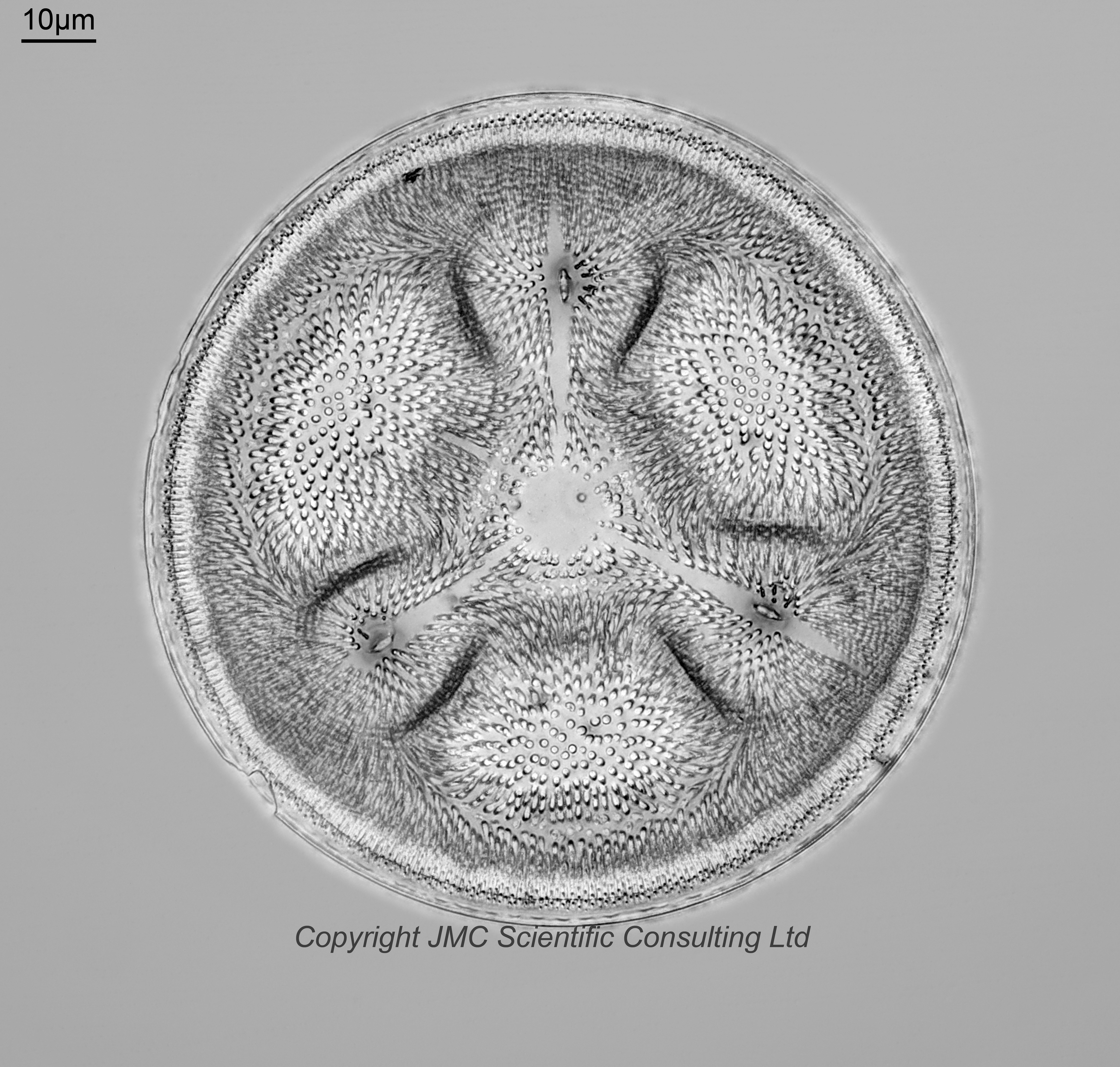
Aulacodiscus subexcavatus. This is a probable ID, although it could be A. excavatus. Brightfield image. Viewed from the underside. 70 images in Pmax.
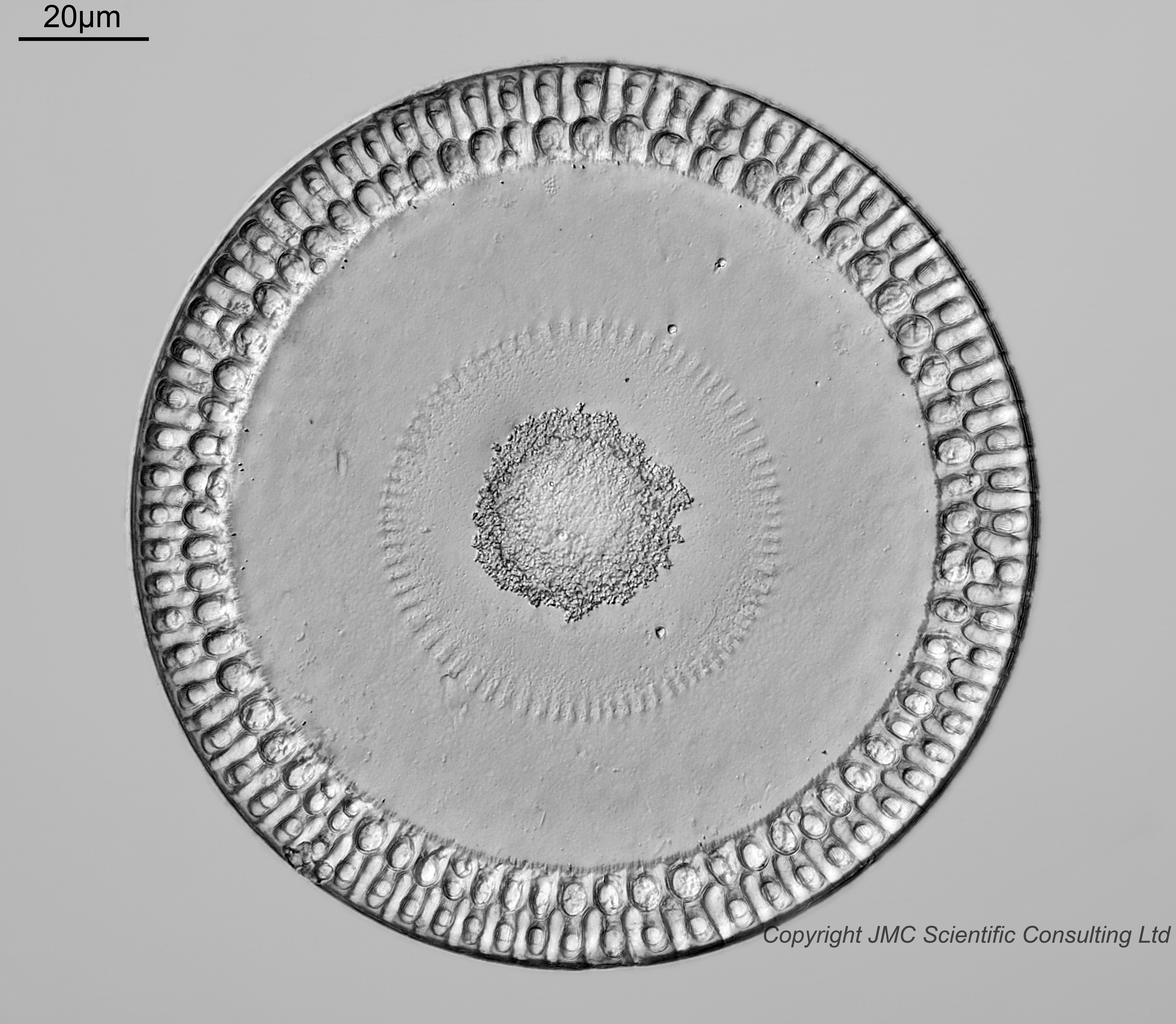
Paralia siberica ? Fairly sure on this one, but not sure as to the variation. Oblique lighting. 34 images in Pmax.
Aulacodiscus sp. (missing a process). Possibly A. subexcavatus. Oblique lighting. 54 images in Pmax. Viewed from above. Aberrant looking form missing a process.
Aulacodiscus archangelskianus. Fairly sure on this one. Brightfield lighting. 48 images in Pmax.
Sheshukovia lustratum ? This is a very tentative ID based on a PhD of ‘Late Cretaceous and Paleocene Marine Diatom Floras’ by Paul Martin Chambers. Another possibility would be Triceratium cellulosum var. simbirskiana (from Schmidt’s Atlas, Plate 112, Figure 4, which is a pretty good match visually). Brightfield lighting. 32 images in Pmax.
Triceratium aculeatum ? Not sure no this one, and this is just a very tentative ID. Mentioned in Greville, R. K. (1861). Descriptions of new and rare Diatoms. Series I. Transactions of the Microscopical Society, New Series, London. 9: 39-45. page(s): p. 45, although no picture given. Brightfield lighting. 33 images in Pmax.
Sheshukovia castellata. Fairly sure on this one. Previous name Triceratium castellatum (and I think for this one that it’s var. fractum). Brightfield lighting. 47 images in Pmax.
Stephanopyxis hannai ? Viewed from the underside. This looks to be a Stephanopyxis sp. of some type and the ID as S. hannai is tentative. Brightfield lighting. 38 images in Pmax.
Entogoniopsis polycistinora. Brightfield lighting. There are 3 examples of this on the slide, and I’ve picked 2 here for imaging.