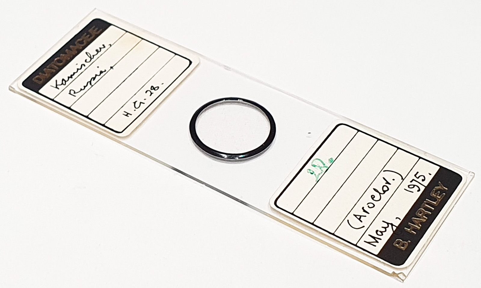
A strew from Kamischev, Russia. Lots of examples of different species present. Prepared by Bernard Hartley. Mounted in Aroclor, and dated May 1975. A relatively tall ring around the coverslip prevented me from using the 63x Leitz Pl Apo objective I’d like to, but I could still use the 40x Leitz Pl Apo instead. Olympus BHB microscope using 450nm LED light. 40x Leitz Pl Apo NA 1.00 objective, oil immersion. Olympus Aplanat Achromat condenser, oil immersion, brightfield lighting. 2.5x Nikon CF PL photoeyepiece. Monochrome converted Nikon d850 camera. Image stacked prepared in Zerene (Pmax).
Various sources used to try and determine the names, including;
Die Diatomeen der Fur-Formation (Alttertiär) aus dem Limfjord-Gebiet, Nordjütland/Dänemark [Diatoms from the Early Tertiary Fur Formation, Limfjord region, Northern Jutland, Denmark], 1991.
Chambers, Paul Martin; (1997) Late Cretaceous and Paleocene marine diatom floras. Doctoral thesis (Ph.D), UCL (University College London).
Some Cretaceous and Palaeogene Trinacria (diatom) Species. Patricia A. Sims, Robert Ross. British Museum (Natural History), 1988.
Nikolaev, V.A., Kociolek, J.P., Fourtanier, E., Barron, J.A. and Harwood, D.M. 2001. Late Cretaceous Diatoms (Bacillariophyceae) from the Marca Shale Member of the Moreno Formation, California. Occasional Papers of the California Academy of Sciences 152:119 pp.
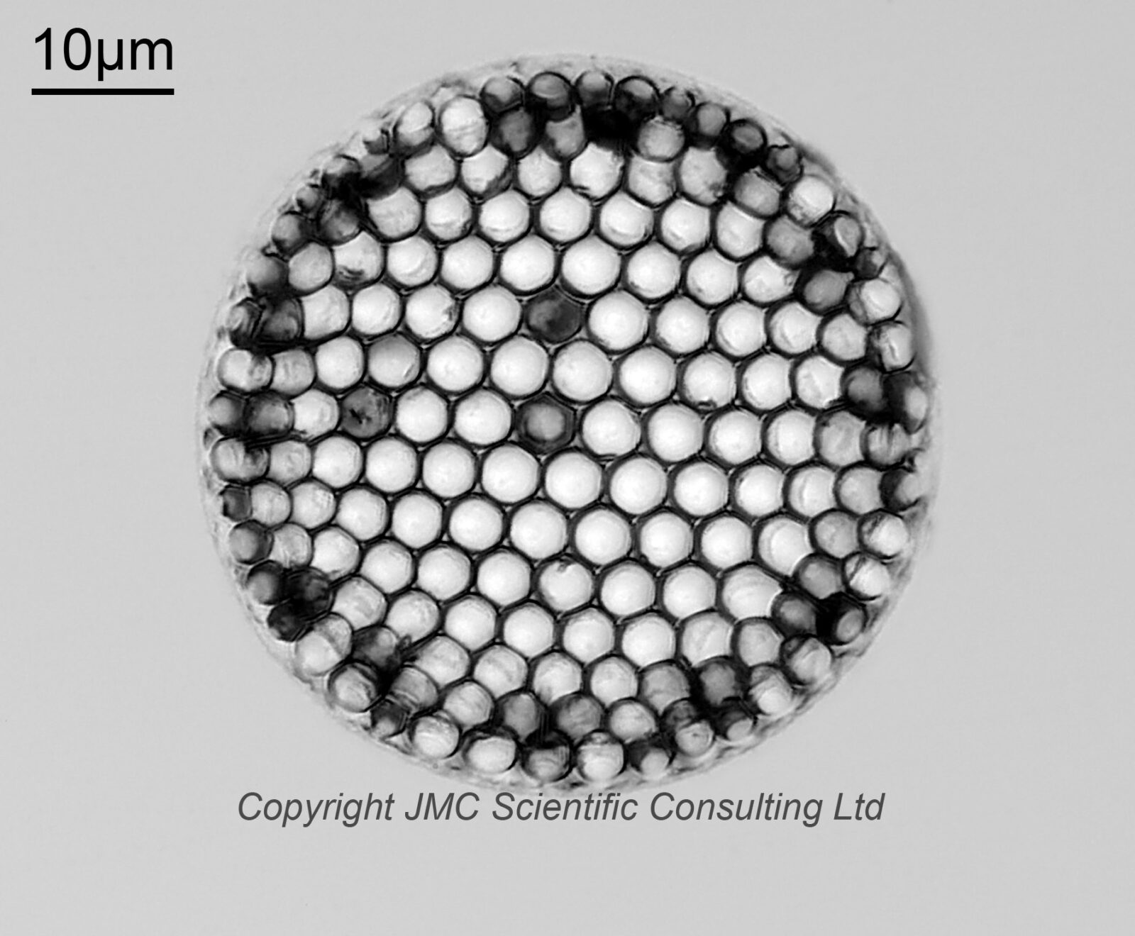
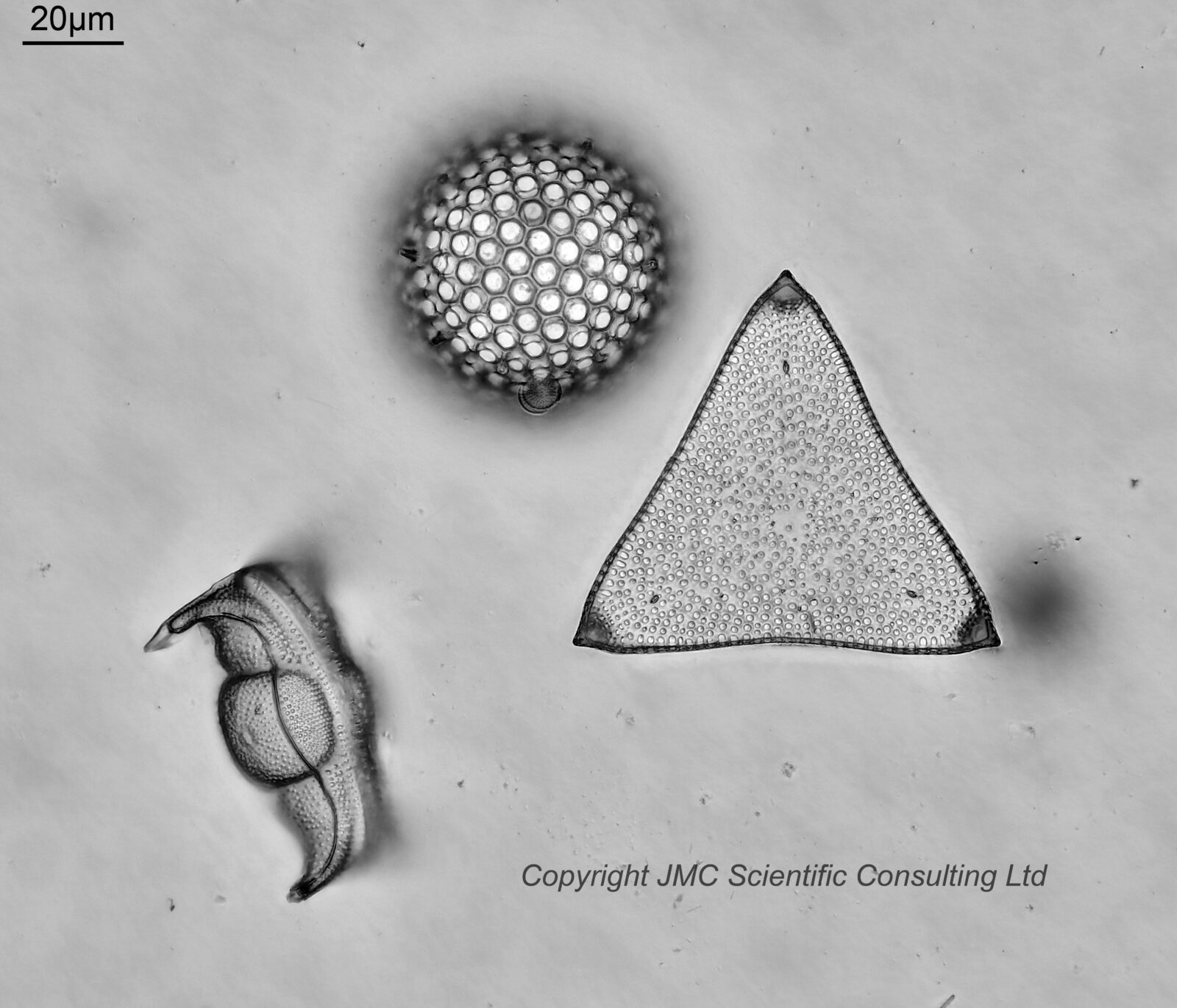
Coscinodiscus sp. (C. marginatus ?). There are quite a few of these with similar structures. Coscinodiscus marginatus seems to be the best match.
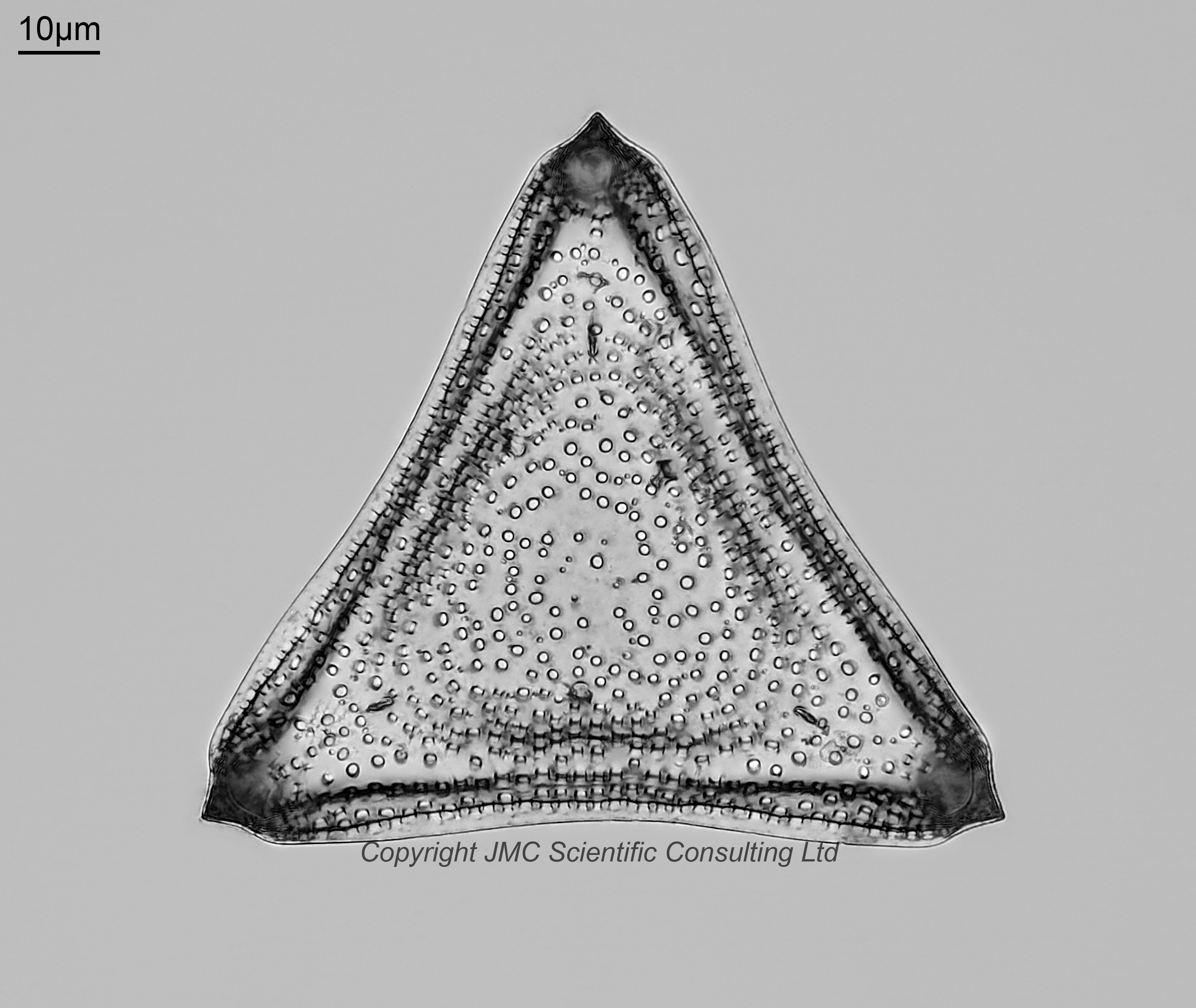
Trinacria excavata. Viewed from the underside. Fairly sure on this one.
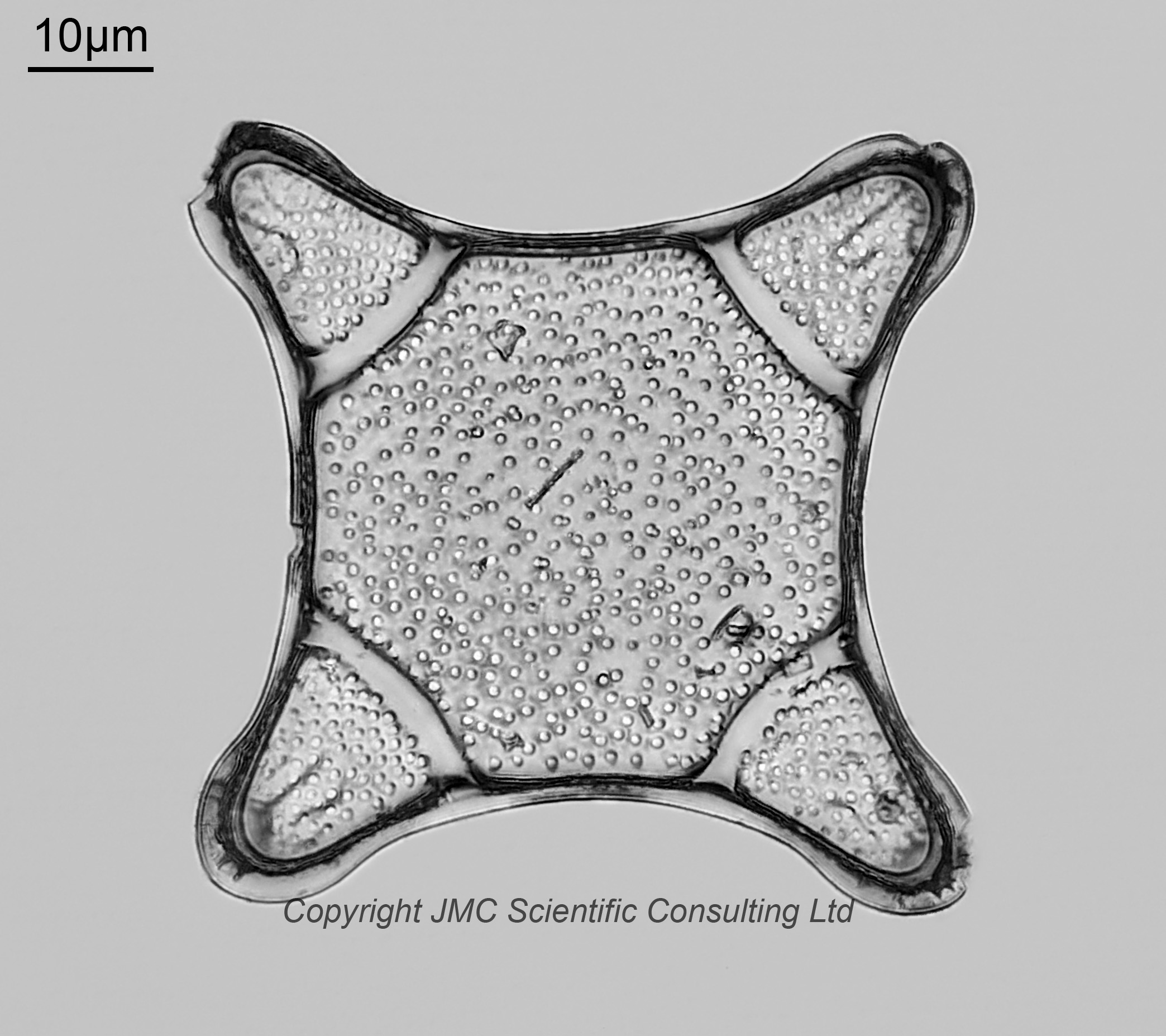
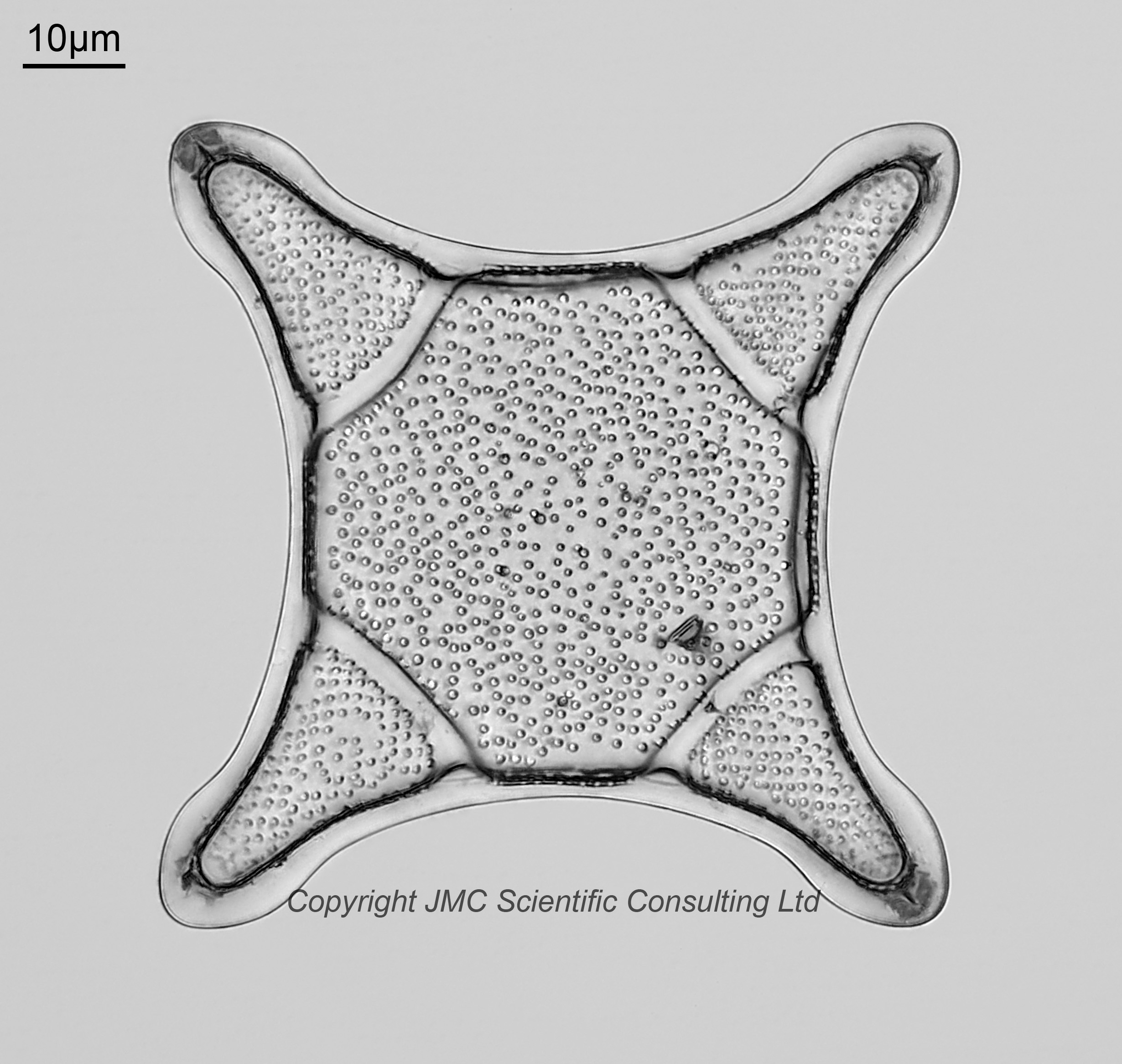
Trinacria regina. Various slightly different shapes and structures, typically viewed from the underside, seems to be in keeping with the variability seen in the Fur Formation book.
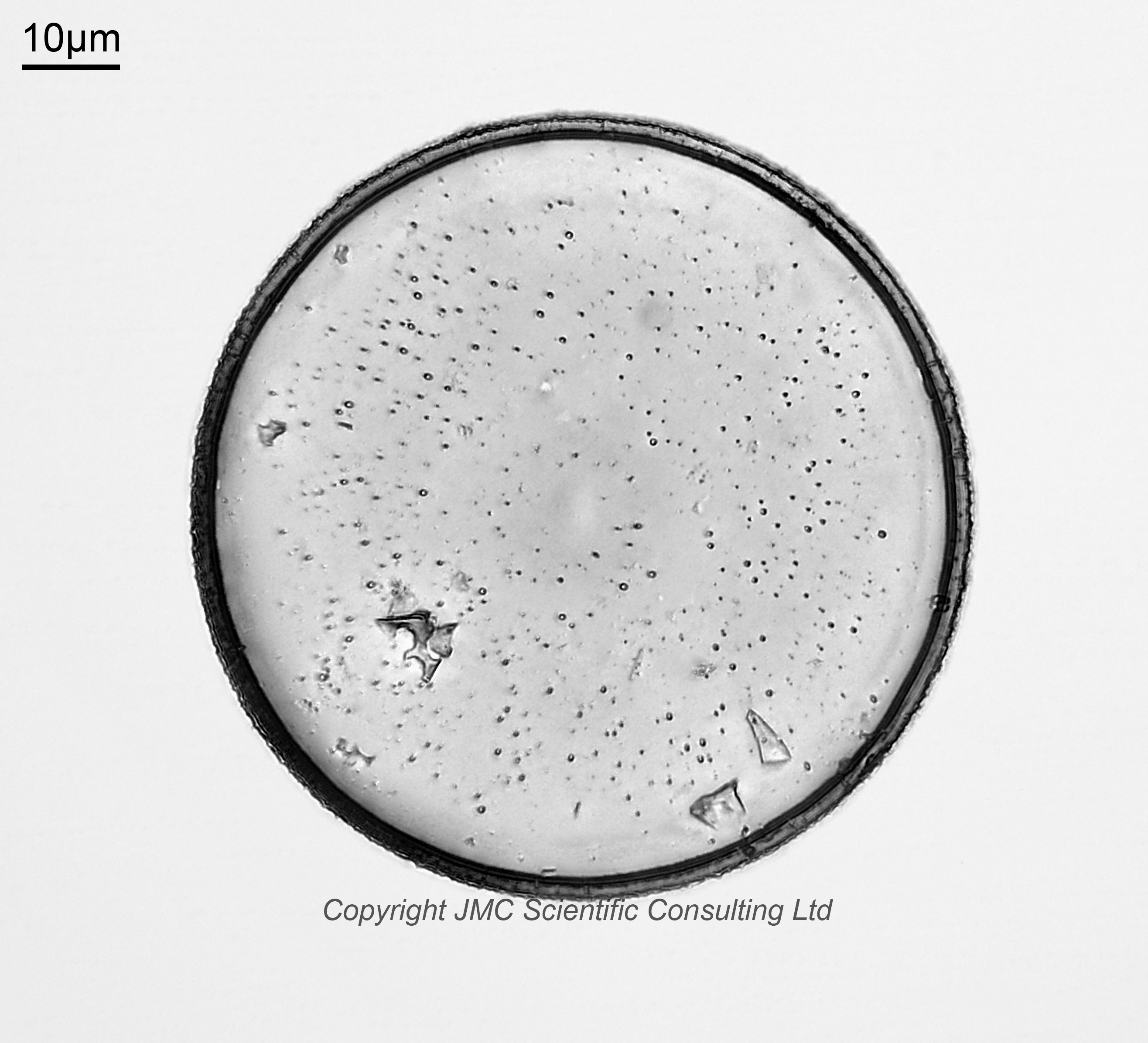
Xanthiopyxis sp. ? Not sure on this one. It looks very much like X. umbonata I have shared on this site (here). Another possibility is Acanthodiscus immaculatus, although that seems to be completely smooth across its surface while this has small bumps there, as such I am less inclined to think it is this.
Solium exsculptum. Both viewed from the underside. Few examples looking slightly different to each other, but in keeping with the examples seen in the Fur Formation book.
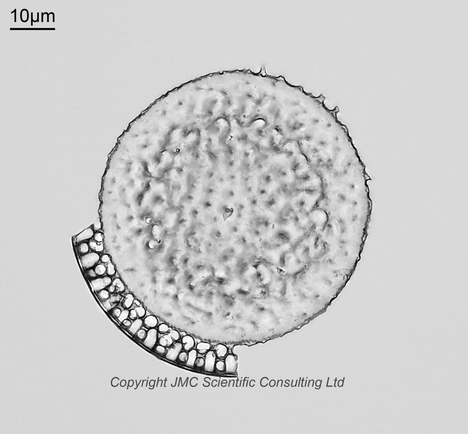
Paralia selecta fragment ? This looks to be a fragment of a diatom. Initially thought it was a Paralia sulcata var siberica fragment (accepted name Melosira sulcata f. siberica), based on Fur Formation book (Plate 30, Figures 7-10). However the more I looked at it the more it looked like Paralia selecta. Published in: Makarova, I.V., Glezer, Z.I., Moiseyeva, A.I. & Nikolaev, V.A. (Eds) (1992). Diatomovyye vodorosli SSSR. Iskopayemyye i sovremennyye [The diatoms of the USSR. Fossil and recent]. Vol. 2(2) pp. 125, 68 plates. Saint Petersburg: Izdatel’stvo Akademii Nauk. [in Russian], Plate 44, 12. Also in Schmidt’s Atlas Plate 177, Figures 40-42.
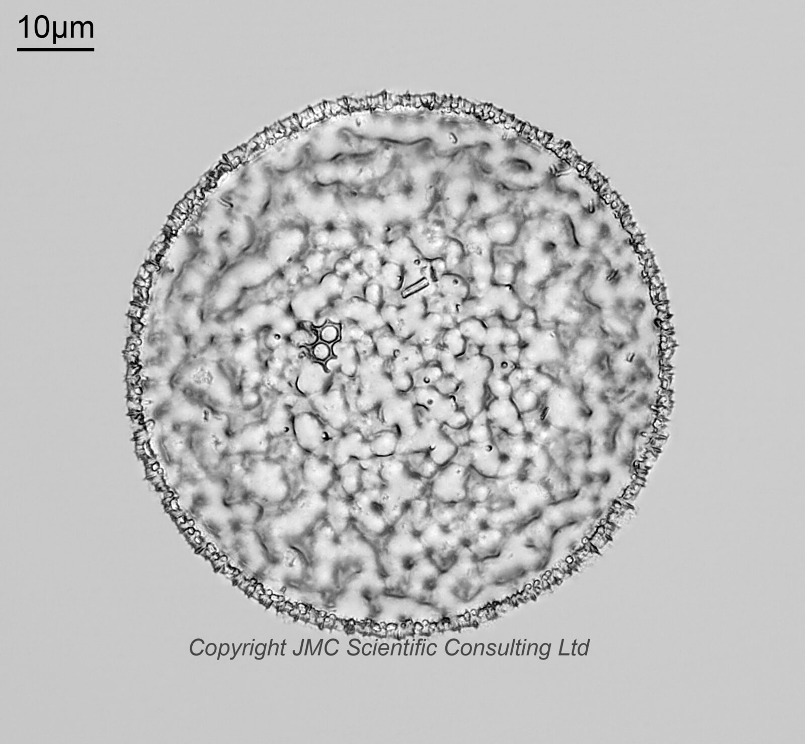
Pseudopodosira sp. ? Not sure on this one. Originally thought that is was similar to the previous one, but without any of the rim, however I think there are differences. The Pseudopodosira sp. identification is based on the Fur Formation book, Plate 54, Fig 10.