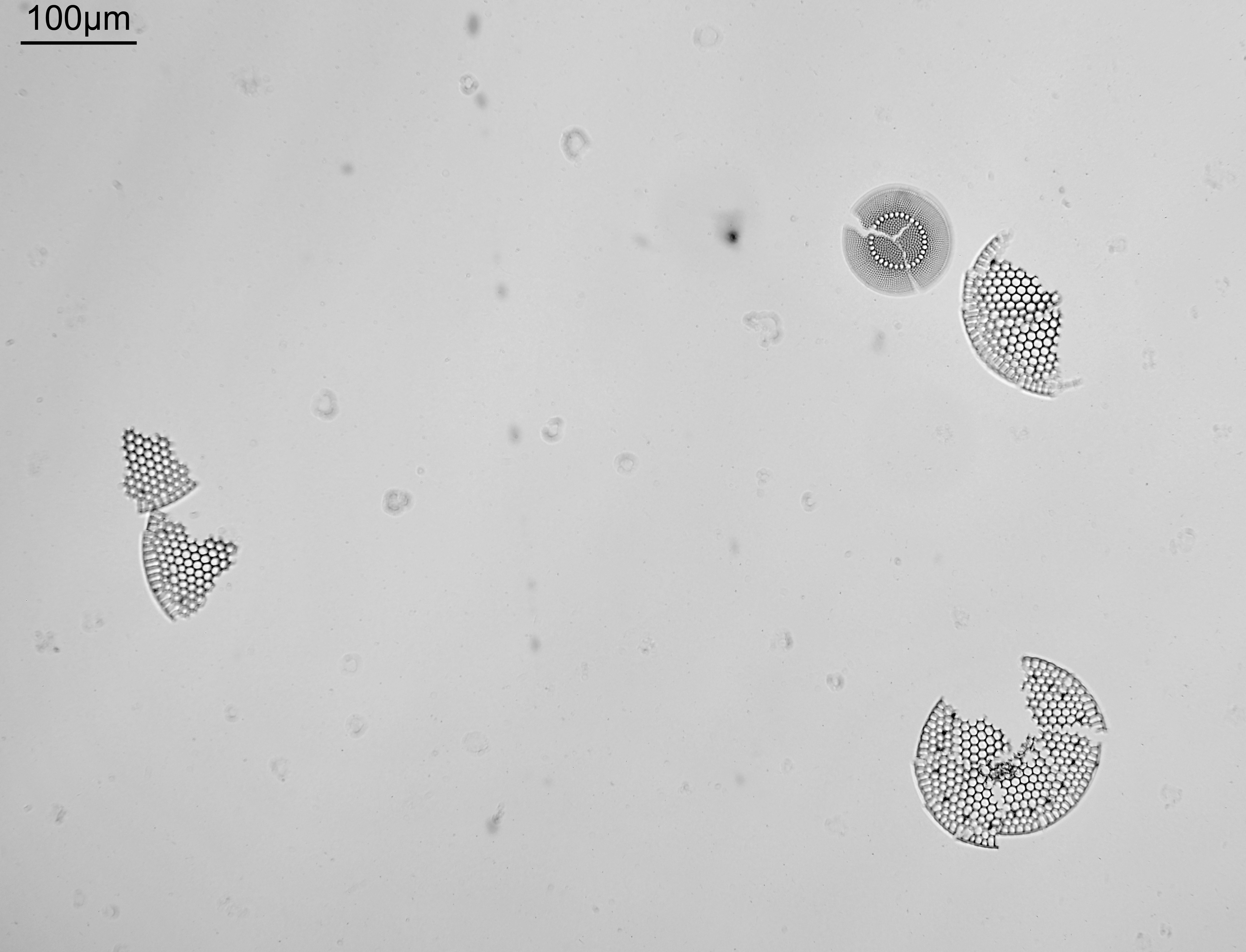
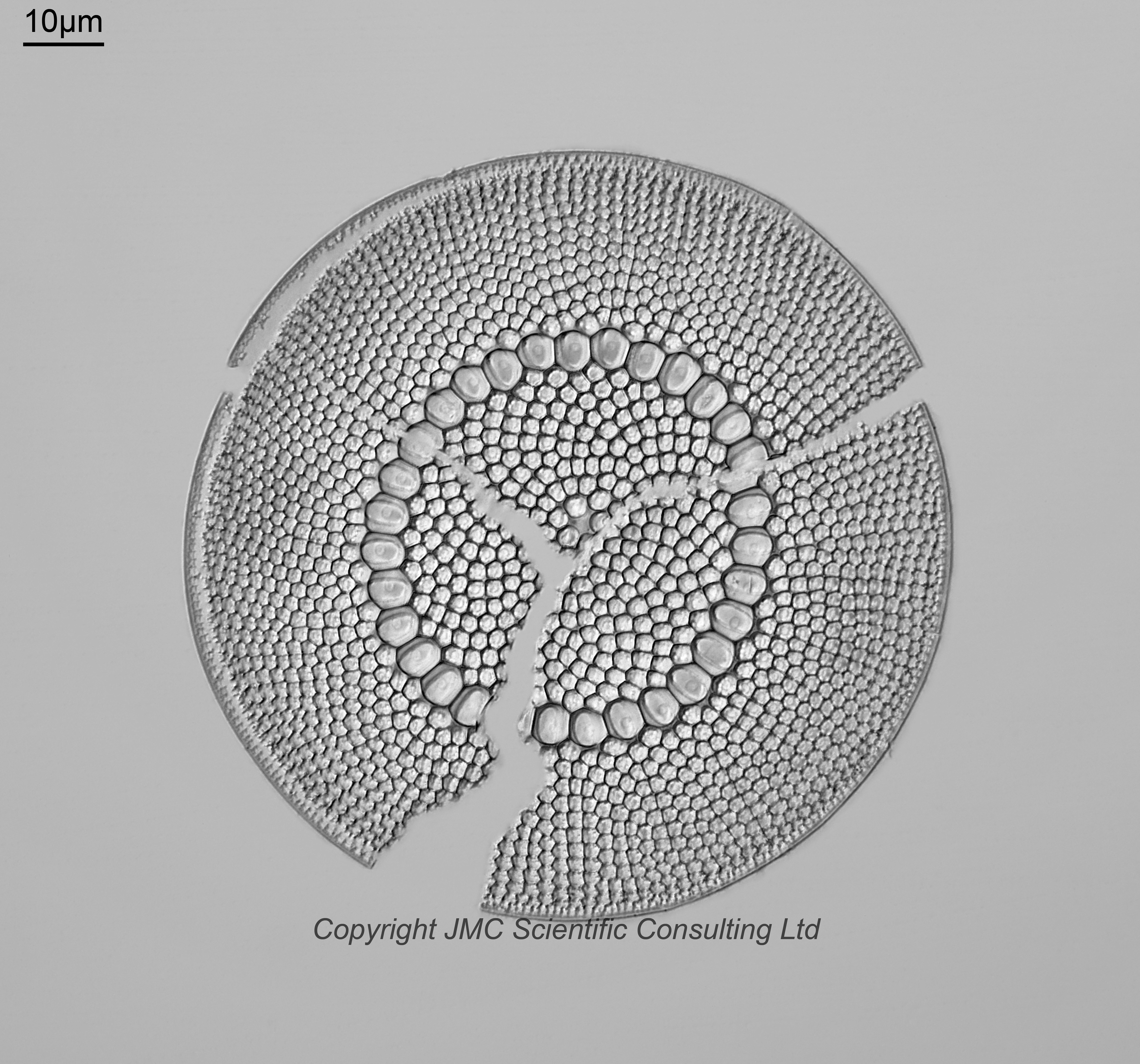
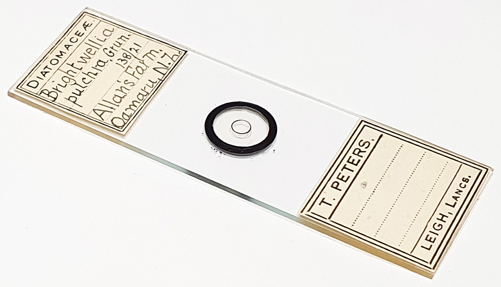
Brightwellia pulchra from Allan’s Farm, Oamaru, New Zealand. Single broken example on the slide mounted with the remains of 2 other diatoms. Prepared by T Peters. There is a reference – 138/21 – referring to Schmidt’s Atlas Plate 138, Figure 21. I suspect this was broken during the mounting stage given the positioning of the pieces. Olympus BHB microscope using 450nm LED light. 63x Leitz Pl Apo 1.4 objective, oil immersion. Olympus Aplanat Achromat condenser, oil immersion, oblique lighting. 2.5x Nikon CF PL photoeyepiece. Monochrome converted Nikon d850 camera. 34 images stacked in Zerene (Pmax).
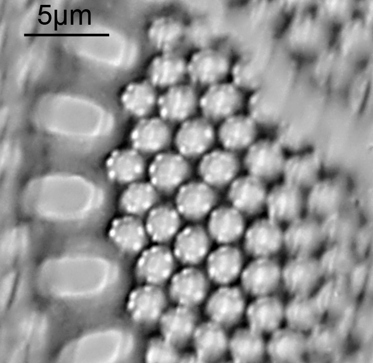
There is an array of bright features around each areolae. These are visible in the final stacked image, but are easier to see in a crop of one of the original images that went into the stack. I didn’t see these in the other example of B. pulchra I have on this site (here), even though the lighting conditions were similar.