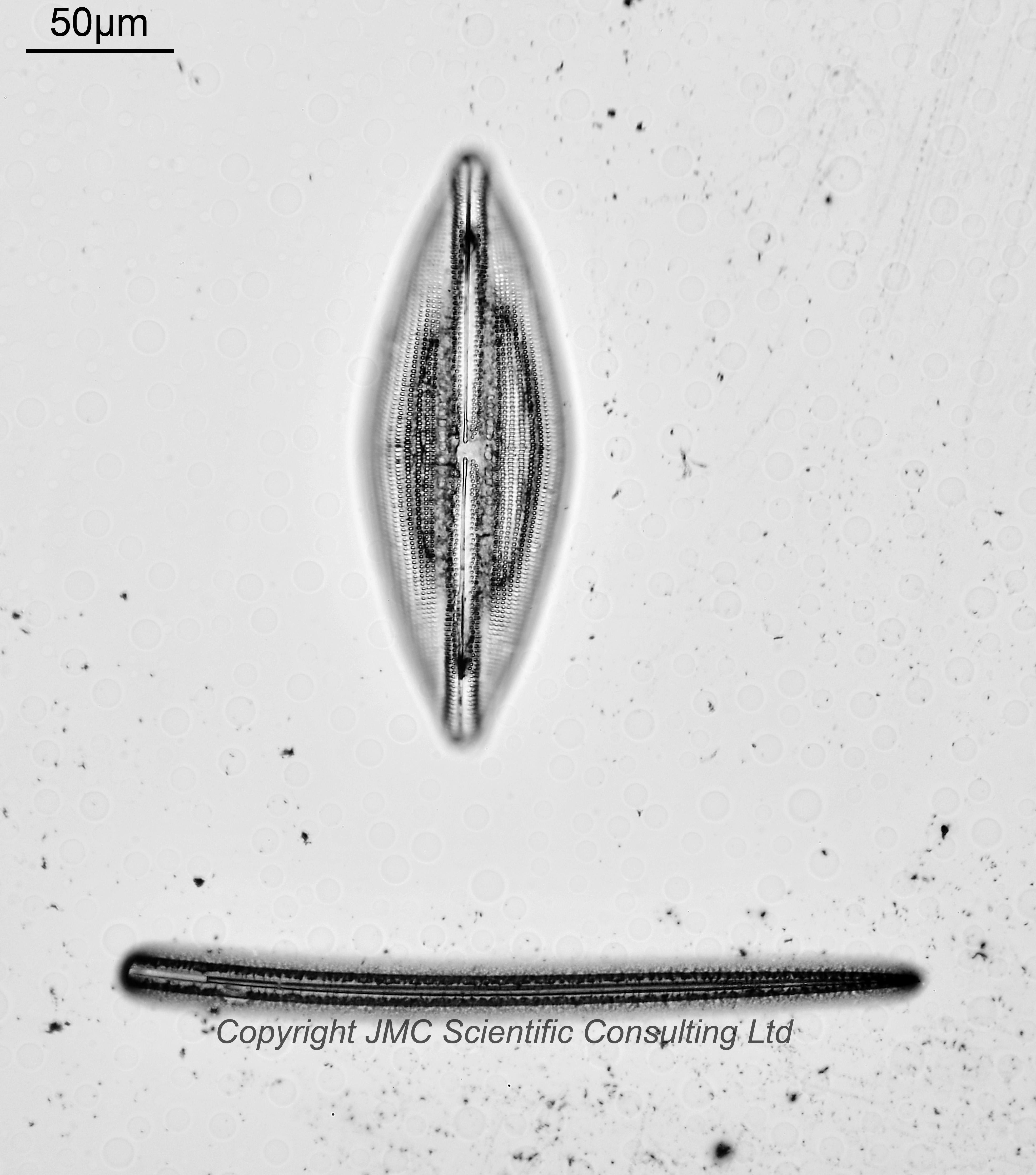
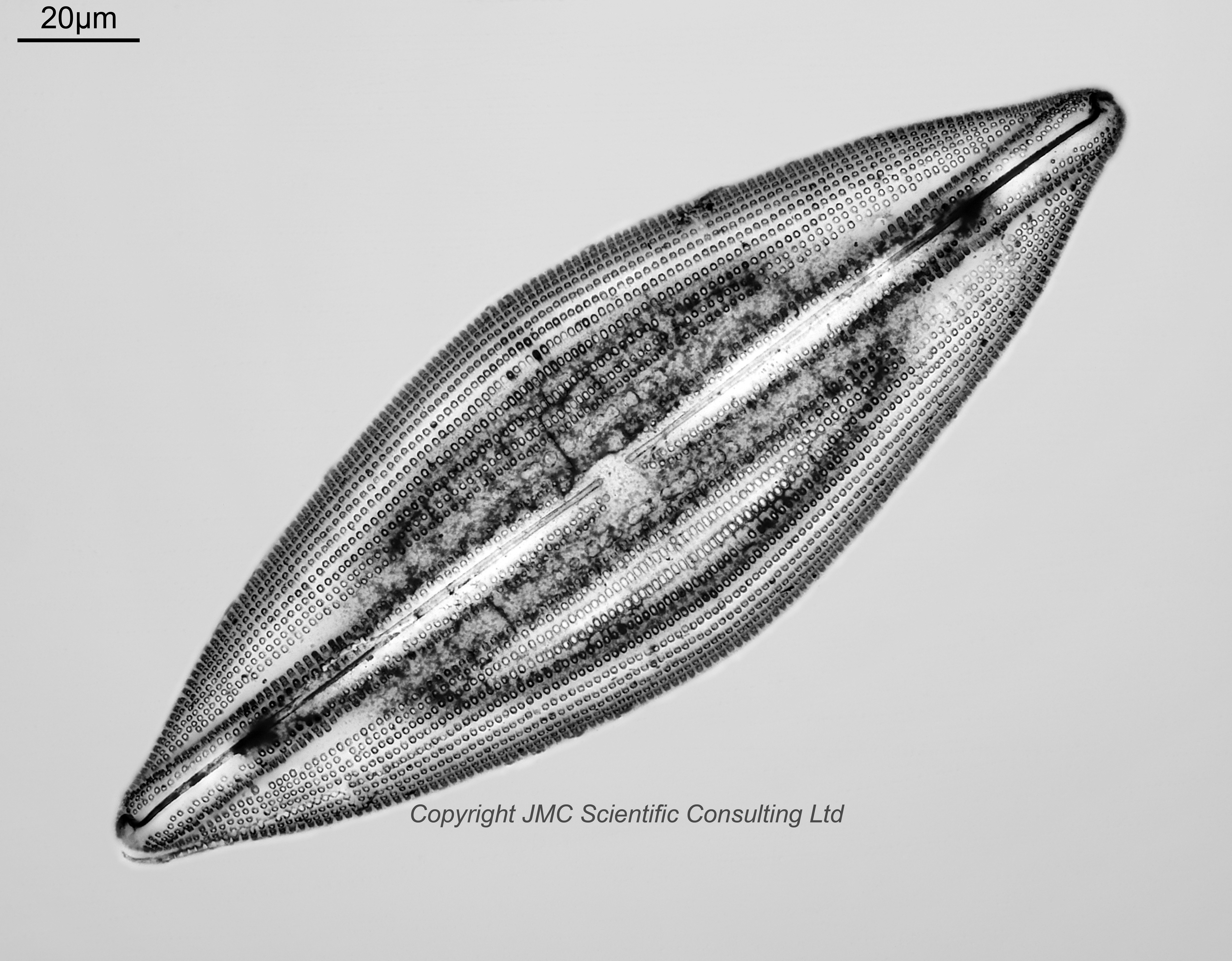
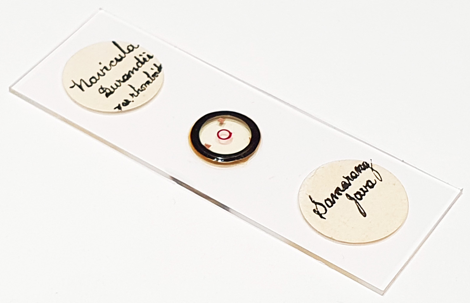
Navicula durandii var. rhomboides from Semarang (spelt Samarang on the slide), Java. Single example on the slide along with a sponge spicule. Prepared by JA Long. Olympus BHB camera using 450nm LED lighting. 63x Leitz Pl Apo NA 1.40 objective, oil immersion. Olympus Aplanat Achromat condenser, oil immersion, brightfield lighting. 2.5x Nikon CF PL photoeyepiece. Monochrome converted Nikon d850 camera. 48 images stacked in Zerene (Pmax).
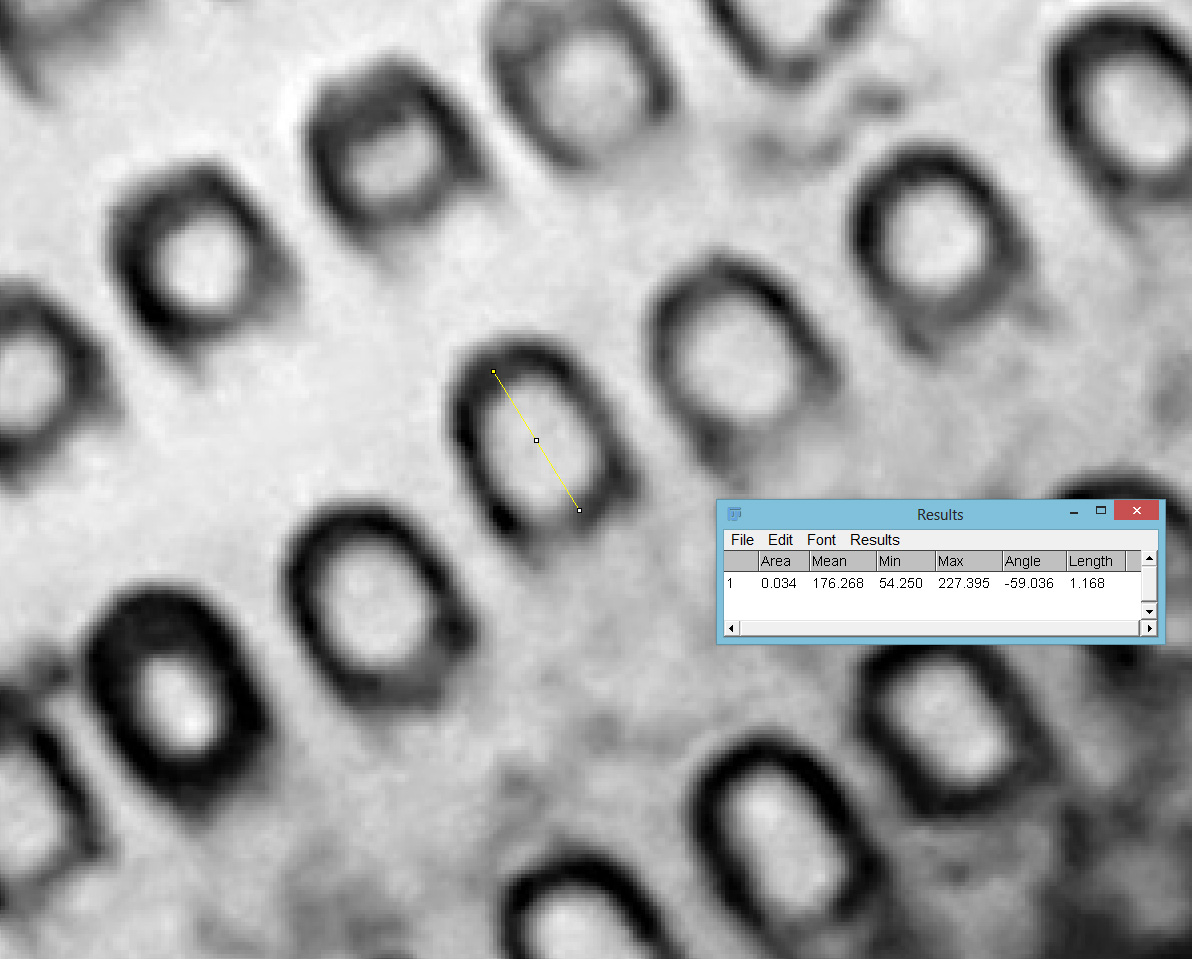
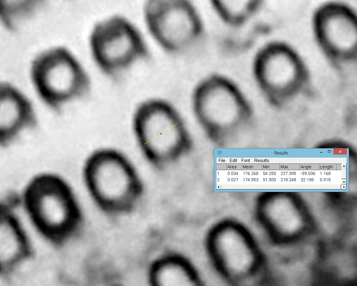
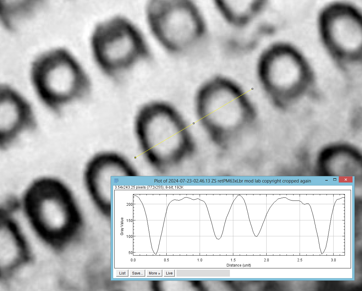
This was an interesting slide to image, and was extremely high contrast, with quite a lot of black material in the background and on the diatom. I did some analysis of the stacked image in ImageJ to have a look at the pores in the structure. Of the one measured, the length was 1168nm and the width 918nm. What was really interesting was the ‘wall thickness’ of the pore. From looking at a transect across a couple of the pores, the wall thickness looked to be around 220-250nm, based on the full width, half max measurement. While above the theoretical resolution of this objective using 450nm light (450/2*1.4 = 161nm) this is getting close to it, and I think down to the nice high contrast image.