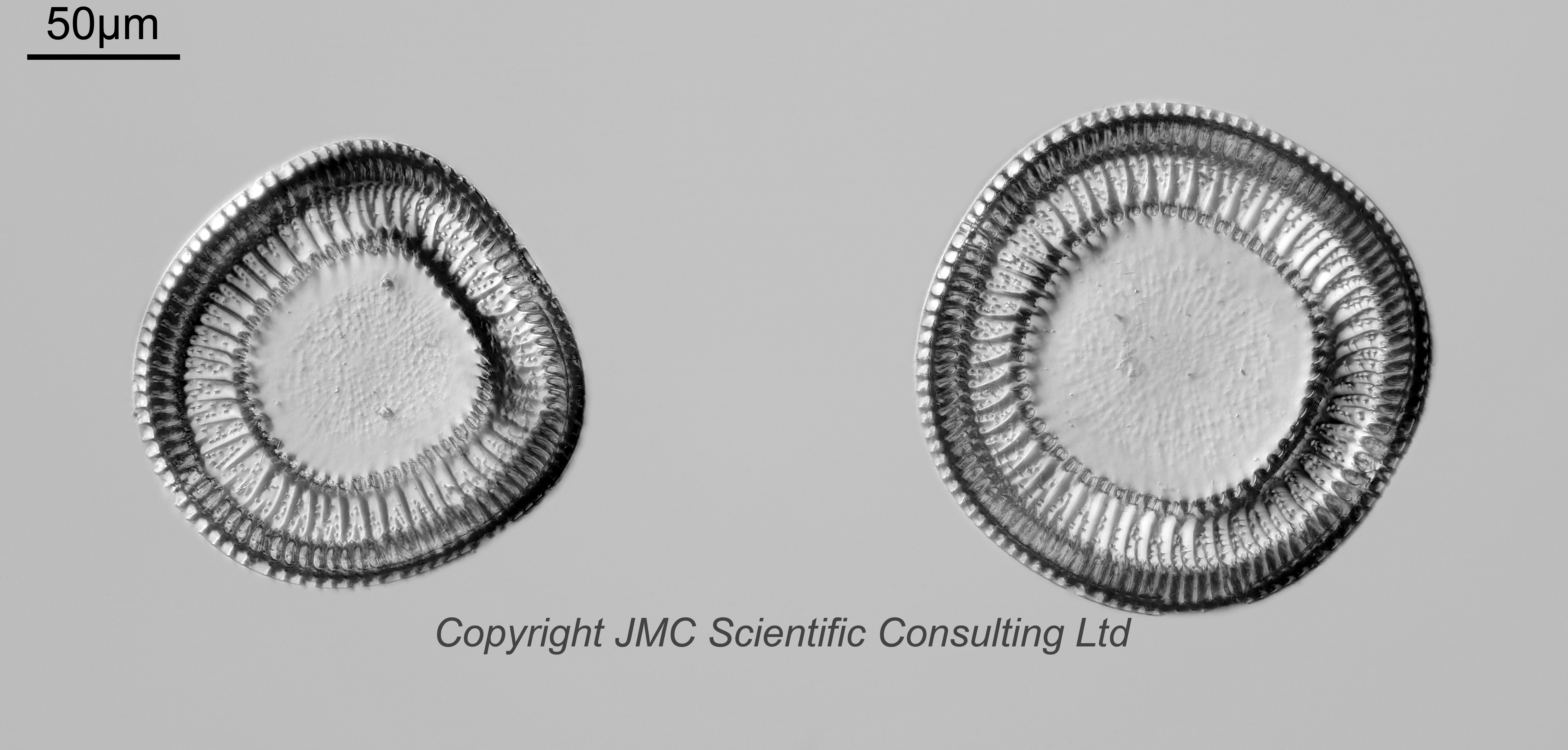
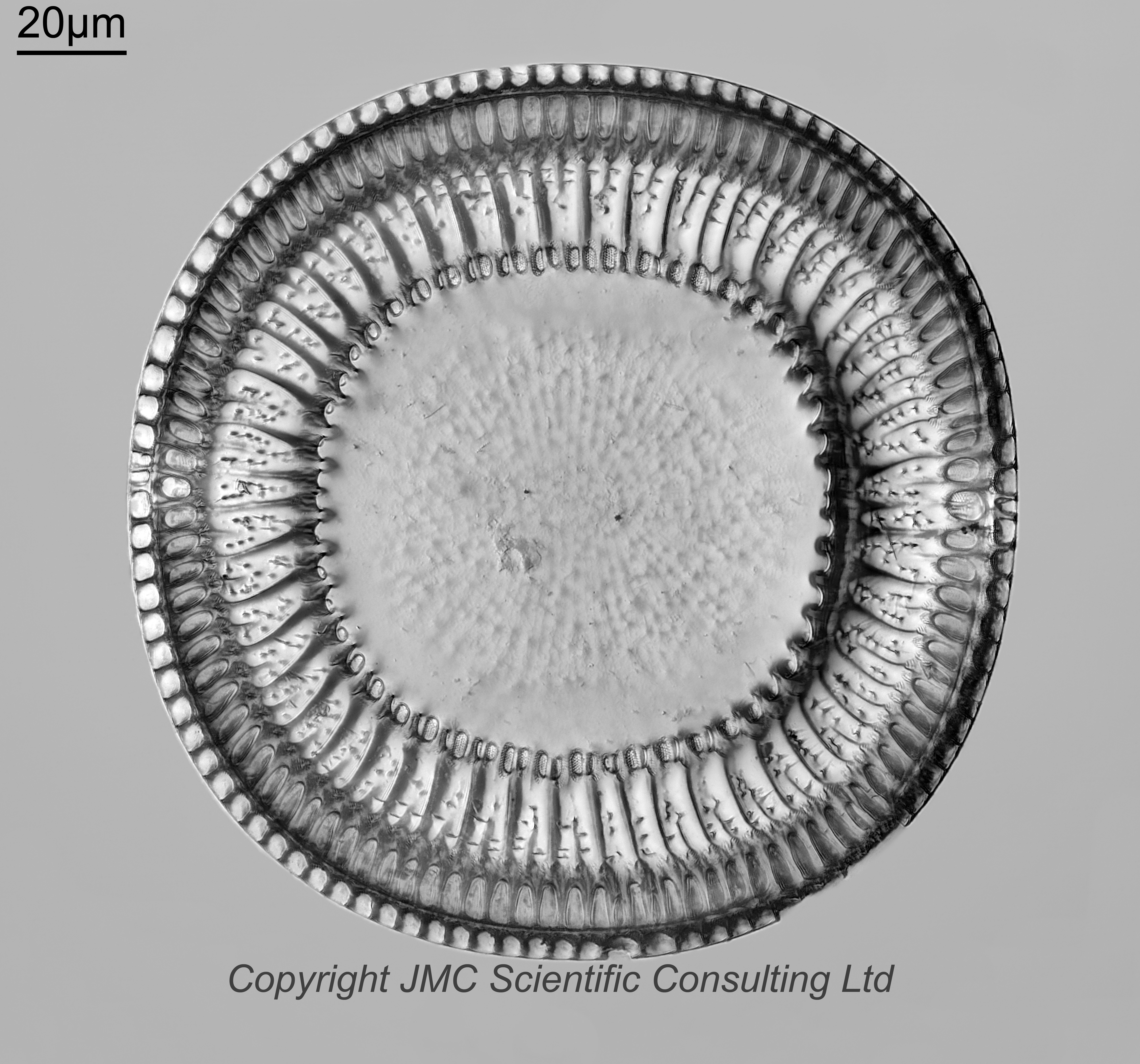
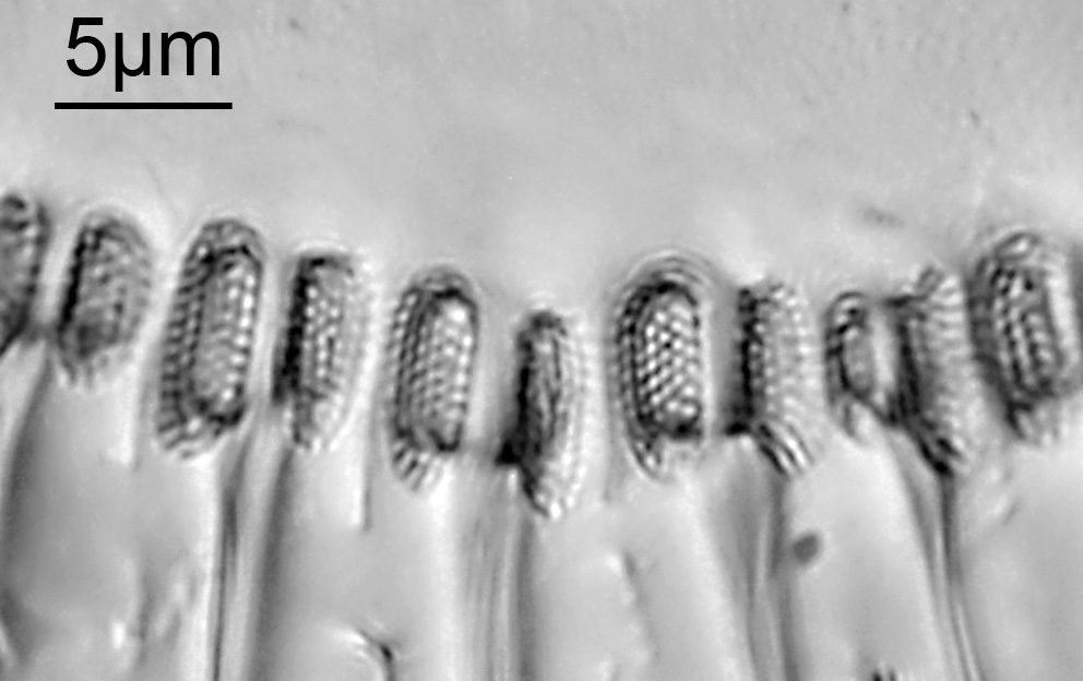
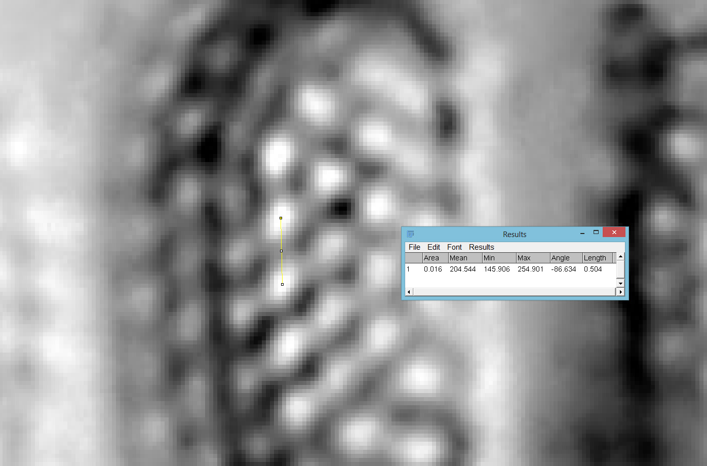
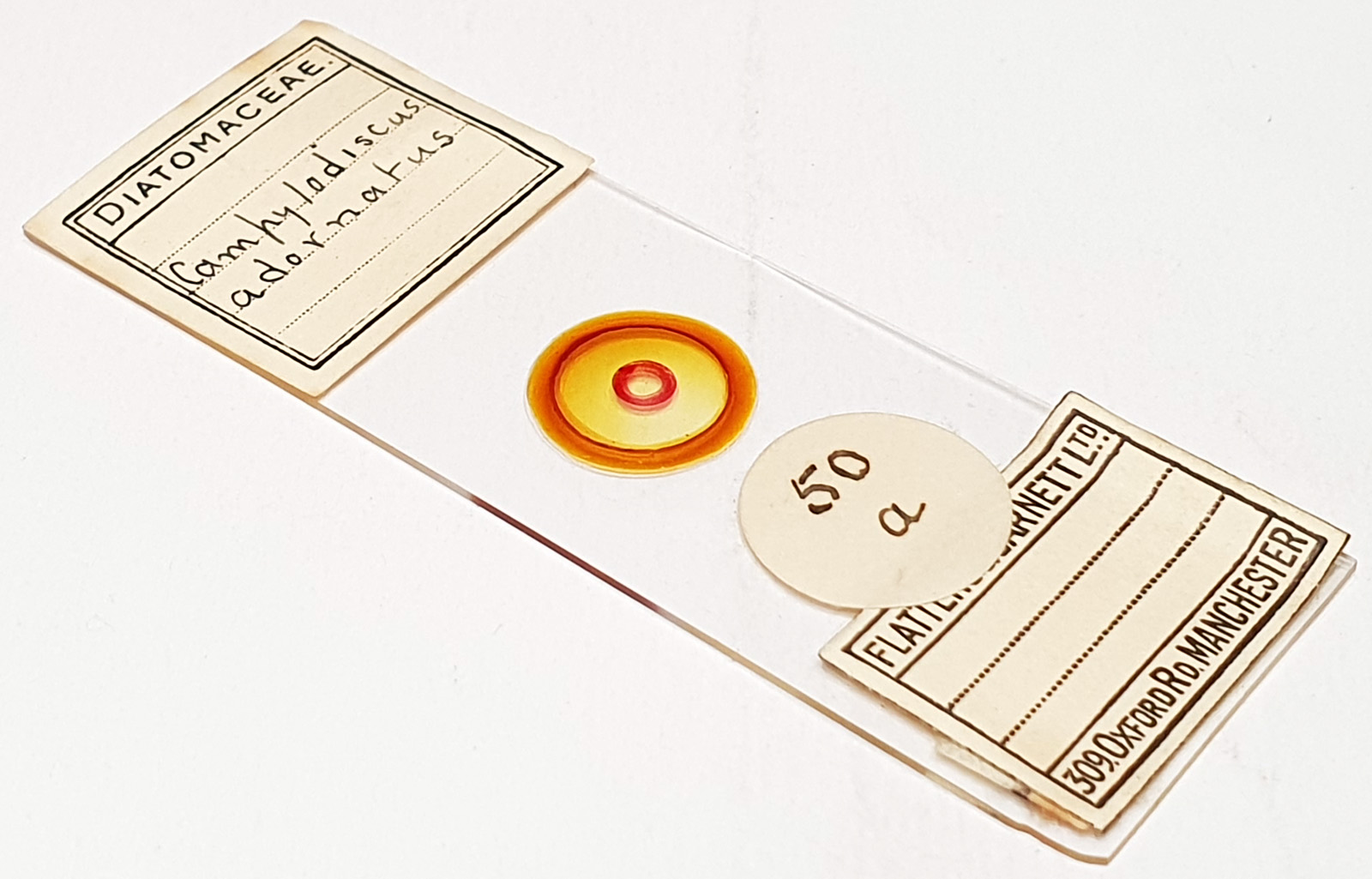
Slide labelled as Campylodiscus adornatus (2 examples). High contrast slide, with some yellowing of the mountant. Prepared by Flatters and Garnett Ltd. Olympus BHB microscope using 450nm LED light. As well as the arrangement I imaged the right hand one using the 63x Leitz Pl Apo 1.4 objective, oil immersion. Olympus Aplanat Achromat condenser, oil immersion, oblique lighting. 2.5x Nikon CF PL photoeyepiece. 11mm removed from the tube the camera to increase the field of view. Monochrome converted Nikon d850 camera. 104 images stacked in Zerene (Pmax). Some amazing details on parts of it, as shown by a crop from one of the original frames. Dot pattern measured as 504nm in ImageJ (middle to middle).
Campylodiscus adornatus A.W.F.Schmidt 1877. Published in: Schmidt, A.[W.F.] (1877). Atlas der Diatomaceen-kunde Series II: Heft 13. pp. pls 49-52. Aschersleben: Verlag von Ernst Schlegel. Plate 51, Figure 5.
In Mann, A. (1925). Marine diatoms of the Philippine Islands. Bulletin United States National Museum 100(6, part 1): 1-182, 39 pls. Page 48, he states “This is rather too close to C. ornatus Greville to justify a separate name.”. Campylodiscus ornatus Greville 1863. Published in: Greville, R.K. (1863). Descriptions of new and rare Diatoms. Series VIII. Transactions of the Microscopical Society, New Series, London 11(3): 13-21, 1 pl. Page 13, Plate I [1], Figure 3.
I’d say these are actually closer to the images of Campylodiscus ornatus according to Greville 1863, and Deby, J., 1891, Analysis of the Diatomaceous Genus Campylodiscus. Privately published, London. However as Mann stated these might be thought of as the same species anyway. Either way, a spectacular Campylodiscus sp..